In vivo imaging method visualizes bone-resorbing cell function in real time
Researchers at at the Immunology Frontier Research Center (IFReC) at Osaka University (Japan) have discovered a way to visualize sites where osteoclasts (bone-resorbing cells) were in the process of resorbing bone in vivo. This real-time visualization of changes in osteoclast localization and activity allowed measurement of bone resorption intensity, and the work could someday contribute to early diagnosis of affected areas and the development of new therapeutic drugs.
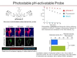
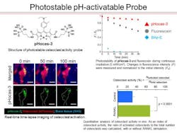
Osteoclasts are bone-resorbing cells that may cause osteoporosis and rheumatoid arthritis if there is an excess in bone resorption. So far, information on osteoclast localization could be gathered by using fluorescent proteins, but this method did not allow an examination of osteoclast activity. So the researchers, led by Kazuya Kikuchi, professor at the Graduate School of Engineering, and Masaru Ishii, professor at the Graduate School of Medicine, produced fluorescent probes that visualize those sites where osteoclasts are in the process of resorbing bone and developed their own imaging device. They thereby conducted an in vivo evaluation of osteoclast function.
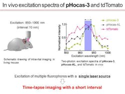
The research team developed a mechanism by which small molecular probes (SMPs) are selectively delivered to the location of target cells. Up until then, application of SMPs to in vivo imaging was particularly challenging, as delivery to target tissues proved difficult. The researchers managed to optimize molecular delivery so that the molecular probes only had to be injected into the mice for imaging. The SMPs were equipped with a switch function that is only triggered at those areas where bone is being resorbed. This enabled the selective visualization of osteoclast activity. In combination with fluorescent proteins that label target cells, the researchers succeeded in real-time visualization of changes in cell localization and activity as well as the measurement of bone resorption intensity.
This research will have great impact in the field of in vivo imaging, as it established a method that allows simple and quick measurement in detecting osteoclasts that are in the process of resorbing bone. This could also benefit early diagnosis and the screening of new therapeutic drugs. The findings also could be applied to molecular design based on physical chemistry principles, creation of functional fluorescent probes by synthetic organic chemistry, and clarification of intravital mechanisms using immunological knowledge and technology.
Full details of the work appear in the journal Nature Chemical Biology; for more information, please visit http://dx.doi.org/10.1038/nchembio.2096.


