OCT, fluorescence imaging pair to better identify heart attack-prone coronary plaques
Researchers from the Wellman Center for Photomedicine at Massachusetts General Hospital (MGH; Boston, MA) have developed a catheter-based device that combines both optical coherence tomography (OCT) and near-infrared autofluorescence (NIRAF) imaging. The method may more accurately identify coronary artery plaques that are most likely to rupture and cause a heart attack.
Related: High-resolution micro-OCT uncovers correlations
"OCT provides images of tissue microstructure, but not of its chemical and molecular composition," says Gary Tearney, MD, Ph.D., of the Wellman Center and the MGH Pathology Department, who is co-senior author of the paper. "Since both of those characteristics are needed to fully understand coronary artery disease, the combination of OCT with NIRAF could provide a more powerful tool for investigating coronary pathology."
The detailed images provided by OCT are created by bouncing NIR light off the internal surfaces of blood vessels, and can identify plaques that have the appearance of rupture-prone "vulnerable" plaques with the potential to cause a heart attack or sudden cardiac death. Fluorescence imaging techniques like NIRAF illuminate an artery with a specific wavelength of light to excite certain molecules, which respond by emitting different wavelengths. Since only certain molecules respond, the resulting signal provides information on the molecular composition of analyzed tissue.
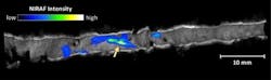
Tearney's team has been investigating whether the additional data provided by NIRAF could identify rupture-prone sites within arterial plaques. Of particular note are fibroatheromas--advanced lesions consisting of a core of dead cells covered by an often-thin fibrous cap, which are particularly prone to rupture. In a previous study using coronary artery segments from cadavers, the research team showed that the NIRAF signal was elevated in fibroatheromas and highest in those with thin fibrous caps, while the current study is investigating the use of NIRAF in living patients.
The study enrolled 12 patients receiving cardiac catheterization at MGH between July 2014 and January 2015. In addition to the clinical procedures conducted to diagnose and/or treat the patients' cardiac disease, Farouc Jaffer, MD, Ph.D., director of MGH Coronary Intervention and co-senior author of the paper, used the novel device developed by the Wellman/MGH research team that acquires both OCT and NIRAF data to construct images of coronary arterial segments. The investigational procedure was identical to that used for conventional OCT imaging.

"Performing OCT-NIRAF imaging is just like conducting standalone coronary OCT imaging, and we are now able to obtain near-infrared fluorescence biological plaque information seamlessly integrated with OCT anatomical images, with no additional time required," Jaffer says. "The clinical success of OCT-NIRAF should further pave the way forward for targeted near-infrared fluorescence molecular imaging using injectable molecular- or cellular-specific agents."
The primary results of the study were confirmation that the procedure was as safe and as feasible to perform as conventional OCT. The OCT-NIRAF images revealed that the NIRAF signal was elevated in areas in which OCT results suggested the presence of a fibroatheroma, and even higher in lesions with thin caps or at sites of plaque rupture and clot formation. Several aspects of the NIRAF signal were different from the patterns produced by other coronary vascular imaging modalities, and more investigation is needed to determine the molecular underpinnings and clinical significance of NIRAF signal results. NIRAF was also elevated in sites showing evidence of inflammation, another potential biomarker of plaques likely to rupture.
"Overall, we believe that the combined OCT-NIRAF examination provides information on molecules within arterial plaques and other features associated with a higher risk of an acute coronary event," says Tearney, a professor of Pathology at Harvard Medical School and the Mike and Sue Hazard Family MGH Research Scholar. "But right now this is a hypothesis, and our findings need to be borne out in larger studies, which we plan to have underway later this year."
Full details of the work appear in the Journal of the American College of Cardiology; for more information, please visit http://dx.doi.org/10.1016/j.jcmg.2015.11.020.