Silver nanoclusters inside synthetic DNA have utility in bioimaging
A team of researchers at the University of Calfornia, Santa Barbara (UCSB) has created nanoscale silver clusters with unique fluorescence properties important for bioimaging.
Related: Better fluorescing, brighter nanophotonics improve diagnostics
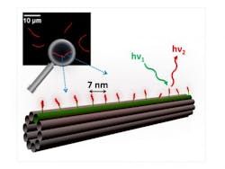
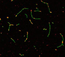
The researchers positioned silver clusters at programmed sites on a nanoscale breadboard, a construction base for prototyping of photonics and electronics. "Our 'breadboard' is a DNA nanotube with spaces programmed 7 nanometers apart," says lead author Stacy Copp, a graduate student in UCSB's Department of Physics.
"Due to the strong interactions between DNA and metal atoms, it's quite challenging to design DNA breadboards that keep their desired structure when these new interactions are introduced," says Beth Gwinn, a professor in UCSB's Department of Physics. "Stacy's work has shown that not only can the breadboard keep its shape when silver clusters are present, it can also position arrays of many hundreds of clusters containing identical numbers of silver atoms—a remarkable degree of control that is promising for realizing new types of nanoscale photonics."
The results of this novel form of DNA nanotechnology address the difficulty of achieving uniform particle sizes and shapes. "In order to make photonic arrays using a self-assembly process, you have to be able to program the positions of the clusters you are putting on the array,"Copp explains. "This paper is the first demonstration of this for silver clusters."
The colors of the clusters are largely determined by the DNA sequence that wraps around them and controls their size. To create a positionable silver cluster with DNA-programmed color, the researchers engineered a piece of DNA with two parts: one that wraps around the cluster and the other that attaches to the DNA nanotube. "Sticking out of the nanotube are short DNA strands that act as docking stations for the silver clusters' host strands," Copp explains.
The research group's team of graduate and undergraduate researchers is able to tune the silver clusters to fluoresce in a wide range of colors, from blue-green all the way to the infrared—an important achievement because tissues have windows of high transparency in the infrared. According to Copp, biologists are always looking for better dye molecules or other infrared (IR)-emitting objects to use for imaging through tissue.
"People are already using similar silver cluster technologies to sense mercury ions, small pieces of DNA that are important for human diseases, and a number of other biochemical molecules," Copp says. "But there's a lot more you can learn by putting the silver clusters on a breadboard instead of doing experiments in a test tube. You get more information if you can see an array of different molecules all at the same time."
The modular design presented in this research means that its step-by-step process can be easily generalized to silver clusters of different sizes and to many types of DNA scaffolds. The paper walks readers through the process of creating the DNA that stabilizes silver clusters. This newly outlined protocol offers investigators a new degree of control and flexibility in the rapidly expanding field of nanophotonics.
The overarching theme of Copp's research is to understand how DNA controls the size and shape of the silver clusters themselves and then figure out how to use the fact that these silver clusters are stabilized by DNA to build nanoscale arrays.
"It's challenging because we don't really understand the interactions between silver and DNA just by itself," Copp says. "So part of what I've been doing is using big datasets to create a bank of working sequences that we've published so other scientists can use them. We want to give researchers tools to design these types of structures intelligently instead of just having to guess."
Full details of the work appear in the journal ACS Nano; for more information, please visit http://dx.doi.org/10.1021/nn506322q.
-----
Follow us on Twitter, 'like' us on Facebook, connect with us on Google+, and join our group on LinkedIn


