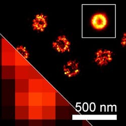
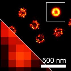
Super-resolution microscopy unravels inner structure of herpes simplex virus
Researchers at the University of Cambridge in England have developed a technique that allows a super-resolution microscopy method called direct stochastic optical reconstruction microscopy (dSTORM) to be used as a structural tool for the study of viruses.
Related: Optical fiber-based sensor IDs smallest virus particles
The work has enabled the research team, led by Prof. Clemens Kaminski and Dr. Colin Crump, to construct an ultra-high-resolution image of herpes simplex virus type 1 (HSV-1) and to determine the position of individual protein layers within the virion with nanometer precision. They also determined the distance between the capsid protein shell and the center of the HSV-1 virion. Additionally, multicolor dSTORM allowed the team to observe multiple layers simultaneously in individual virus particles.
"Pinpointing with nanometric precision the position of individual proteins within the HSV-1 structure is crucial in providing the research community with potential therapeutic targets," says Dr. Romain Laine of the Department of Chemical Engineering and Biotechnology's Laser Analytics Group at the University of Cambridge. "Understanding the structure gives us a chance to better understand the functions of the different proteins present in the virus, even to suggest new functionality, and to consider previously unforeseeable protein-protein interactions."
Full details of the work appear in the journal Nature Communications; for more information, please visit http://dx.doi.org/10.1038/ncomms6980.
-----
Follow us on Twitter, 'like' us on Facebook, connect with us on Google+, and join our group on LinkedIn

