Multimodal fluorescence imaging method peers into nanoscale pores for use in drug development
A team of researchers at Rice University (Houston, TX), along with collaborators at the University of California, Los Angeles (UCLA) and Kansas State University (Manhattan, KS), has developed a multimodal fluorescence imaging method that can peer into and measure the space in porous materials, even if that space is too small or fragile for traditional microscopes.
Related: Imaging system combines five molecular imaging techniques
The Rice University team, led by chemist Christy Landes, invented a technique to characterize such nanoscale spaces, an important advance toward her group’s ongoing project to efficiently separate “proteins of interest” for drug development. It should also benefit the analysis of porous materials of all kinds, like liquid crystals, hydrogels, polymers, and even biological substances like cytosol, the compartmentalized fluids in cells.
Standard techniques like atomic force, x-ray, and electron microscopy would require samples to be either frozen or dried. “That would either shrink or swell or change their structures,” Landes says.
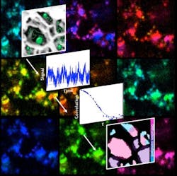
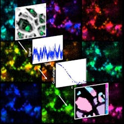
So, Landes and her team combined their experience with super-resolution microscopy and fluorescence correlation spectroscopy techniques. Super-resolution microscopy is a way to see objects at resolutions below the diffraction limit, which restricts the viewing of things that are smaller than the wavelength of light directed at them. Correlation spectroscopy is a way to measure fluorescent particles as they fluctuate. By crunching data collected via a combination of super-resolution microscopy and correlation spectroscopy, the researchers mapped slices of the material to see where charged particles tended to cluster.
The combined method, which they call fluorescence correlation spectroscopy super-resolution optical fluctuation imaging (fcsSOFI), measures fluorescent tags as they diffuse in the pores, which allows researchers to simultaneously characterize dimensions and dynamics within the pores. The lab tested its technique on both soft agarose hydrogels and lyotropic liquid crystals. Next, they plan to extend their mapping to three-dimensional spaces.
“We now have both pieces of our puzzle: We can see our proteins interacting with charges within our porous material, and we can measure the pores,” Landes says. “This has direct relevance to the protein separation problem for the $100 billion pharmaceutical industry.”
Full details of the work appear in the journal ACS Nano; for more information, please visit http://dx.doi.org/10.1021/acsnano.5b03430.
Follow us on Twitter, 'like' us on Facebook, connect with us on Google+, and join our group on LinkedIn

