Flagship Biosciences to integrate color calibration into tissue analysis platform

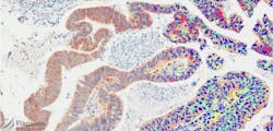
Tissue imaging specialist Flagship Biosciences (Westminster, CO) will integrate Datacolor (Lawrenceville, NJ)'s ChromaCal color calibration technology into its Tissue Analysis Laboratory Information System (TALIS) digital imaging platform.
Related: Advancing microscopy with apps
ChromaCal, a commercially available color calibration technology for brightfield microscopy, addresses the critical need for quality assurance standards in digital microscopy imaging. With the technology, images captured over various time periods and using a variety of automated imaging systems can be standardized to deliver consistency in quality and color, and therefore optimize evaluation and automated analysis. The integration ensures standardization of color parameters, leading to consistency and accuracy of an image’s color base and highly reproducible results across images.
The collaboration allows Flagship Biosciences to include whole-slide imaging systems and scanners in its offering, according to Mark S. McNulty, general manager for Datacolor ChromaCal. For more information, please contact McNulty at [email protected].
Follow us on Twitter, 'like' us on Facebook, connect with us on Google+, and join our group on LinkedIn