MULTIPHOTON MICROSCOPY/FLUORESCENT PROTEINS: New approach overcomes limitations of two-photon dual-color imaging
Two-photon dual-color imaging of tissues and cells labeled with fluorescent proteins (FPs) is a powerful microscopy tool. But the approach is powerfully challenging, too–because most two-photon microscopes are fitted with one (costly) femtosecond laser and thus can provide just one excitation wavelength at a time. And a single excitation wavelength rarely produces well-separated fluorescence emission spectra.
Current methods of two-photon dual-color imaging are limited by the requirement of large differences in Stokes shifts between the FPs used and their low two-photon absorption (2PA) efficiency. Various attempts have been made to overcome this limitation, but their applicability has been limited for various reasons.
A new approach developed by a team of scientists from Montana State University (Bozeman, MT) uses the simultaneous excitation of the lowest-energy electronic transition of a blue fluorescent protein and a higher-energy electronic transition of a red fluorescent protein.1 The method does not require large differences in Stokes shifts and can be extended to a variety of FP pairs with larger 2PA efficiency and more optimal imaging properties.
Characterizing candidates
The Montana State team performed a detailed characterization of the two-photon absorption (2PA) properties of a series of orange and red FPs over a wide range of wavelengths (650–1300 nm). The spectra reveal optimal wavelengths for excitation, new excitable two-photon transitions, and the best absorbing and brightest FPs in the series for use in two-photon laser scanning microscopy (TPLSM).
One of the proteins, tagRFP, has a high two-photon cross-section in the range of 700–780 nm. Two-photon absorption at this range is due to a higher-energy or short-wavelength transition(s) of the tagRFP chromophore, and is within the range of the modelocked Ti:sapphire lasers commonly found on commercially available two-photon microscopes. The team reasoned that if they could identify an FP with a strong lowest-energy, short-wavelength transition in the same 700–780-nm region, it would provide an ideal partner with tagRFP. So they collected 2PA spectra for a series of recently developed blue, teal, and green FPs that are photostable, pH insensitive, have relatively high quantum yields and extinction coefficients, and work well in fusion tags.
The 2PA spectra of all the FPs turned out to be similar in that they consist of two distinct absorption bands. The longer wavelength band is attributed to the lowest-energy electronic (S0→S1) transition while the short wavelength band results from transitions to a higher-energy electronic state(s) (S0→Sn). Partially resolved vibronic transitions give the longer wavelength band its complex structure, and with the exception of mAmetrine, whose 1PA and 2PA maxima coincide, the dominant 2PA peak is blue-shifted compared to the 1PA spectrum due to a vibronic redistribution of intensity.
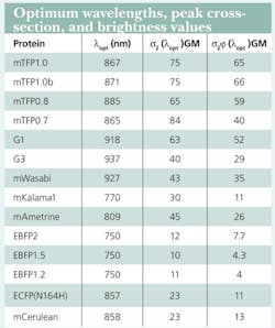
According to the team, the FPs studied have the advantage that the longer-wavelength 2PA band of their chromophores lies within the tuning range of commercial Ti:sapphire lasers commonly used in TPLSM. All exhibit relatively high 2PA efficiency (cross-section, σ2). The FPs in the EBFP series (BFP Y66H chromophore) have very similar cross-section values on the order of 10 GM at ~750 nm (see table).
The spectra reveal that mKalama1 is particularly well suited as a partner with tagRFP in two-photon dual-color imaging. The higher excited state revealed in the tagRFP spectrum makes it possible to couple this bright two-photon probe with mKalama1 such that a single wavelength can excite both optimally. The blue emission of mKalama1 is a limitation to this approach because of potential scattering and absorption in deep tissue imaging applications. This limitation, however, is balanced by the advantages of two-photon excitation and access to multicolor imaging with only one laser. Further, the method of exploiting the different excitable transitions of FPs for simultaneous imaging is highly extensible. One can easily imagine finding FP pairs with favorable properties such as further red-shifted excitations and emissions, greater photostability, and higher two-photon brightness.
References
- Tillo et al., BMC Biotechnol. 2010, 10:6.
About the Author

Barbara Gefvert
Editor-in-Chief, BioOptics World (2008-2020)
Barbara G. Gefvert has been a science and technology editor and writer since 1987, and served as editor in chief on multiple publications, including Sensors magazine for nearly a decade.
