Hybrid imaging approach may expedite personalized medicine
What do you get when you combine coherent anti-Stokes Raman scattering (CARS) and two-photon excited fluorescence (TPEF) microscopy with fluorescence recovery after photobleaching (FRAP)? If you're a researcher at the State University of New York's University at Buffalo (UB), you get a demonstration of the ability to monitor, in real time, the transformations that cellular macromolecules undergo during apoptosis (programmed cell death).1 You also get a potentially valuable tool for screening new drug compounds under development–and the opportunity to help realize the potential of customized molecular medicine, in which chemotherapy, for example, can be precisely targeted to cellular changes exhibited by individual patients.
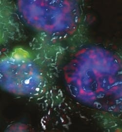
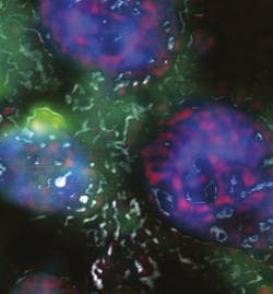
Led by Paras N. Prasad, executive director of the UB Institute for Lasers, Photonics and Biophotonics (ILPB), the interdisciplinary team used the approach to measure dynamics of proteins. The resulting composite imagery integrates into one picture the information on all four types of biomolecules, each type being represented by a different color: proteins in red, RNA in green, DNA in blue, and lipids in grey.
The approach enables "dynamic mapping of the transformations occurring in the cell at the molecular level," says Prasad. "For the first time, this approach allows us to monitor in a single scan, four different types of images, characterizing the distribution of proteins, DNA, RNA and lipids in the cell," adds Aliaksandr V. Kachynski, research associate professor at the ILPB and a co-author of the paper describing it. The distributions change dramatically as cells undergo apoptosis.
The results will facilitate monitoring of how specific cancer drugs affect individual cells throughout treatment, and enable determination of doses so as to minimize side effects.
A new paper from the UB team, forthcoming in Biophysical Journal, further extends this work.
1. A. Pliss et al., PNAS 107 (29): 12771-12776 (2010)
More BioOptics World Current Issue Articles
More BioOptics World Archives Issue Articles

