Nanotechnology is revolutionizing optics in general," says Srinivas Sridhar, a physicist at Northeastern University (Boston, MA). "It lets you make entirely new materials, which have entirely novel properties." For the biological sciences, Sridhar sees two useful applications of nanotechnology. One involves the construction of new optical components, and the other revolves around nano-size particles.
Lenses with focus on fluorescence
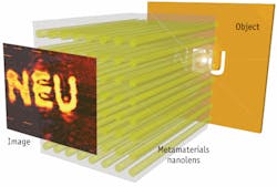
In terms of nano-driven optical components, lenses make up a key area. The size of a lens' components and the working range often drop into nano-scale dimensions. For example, if light bounces off an object at a high enough angle, it creates evanescent waves, which decay in just a few nanometers. A so-called superlens can capture those waves and use them to construct an image with super-high resolution.
Sridhar and his colleagues have demonstrated such a superlens, constructed from an array of nanowires.1 "It can beat the diffraction limit by at least a factor of two," he says.
Other techniques can also turn out superlenses–for instance, a stack of metallic films can make a superlens. Unfortunately for biologists, though, such a superlens runs into trouble with fluorescent dyes, which interact with the metals, thereby reducing the intensity of a fluorescence and increasing its decay rate.
To solve that problem, Kareem Elsayad and Katrin Heinze–both at the optical engineering laboratory at the Research Institute of Molecular Pathology (Vienna, Austria)–turned to computation.2 Using a superlens design of metallic and dielectric layers stacked on top of each other, Elsayad and Heinze calculated the best way to capture light from green fluorescent protein (GFP). The key lies in getting the superlens very close to the specimen–but not too close. For maximum effectiveness, the lens needs to be within 30 nanometers of the dye, but farther than 10 nanometers away from it. So Elsayad says, "Imaging of fluorescently labeled molecules or molecular complexes on cell surfaces such as membrane complexes would be a potential application of the proposed superlens." He adds, "One example would be the adhesion sites in migrating cells. The position of a GFP-labeled adhesion complex protein could be localized beyond the diffraction limit when it is close enough to the superlens surface."Beyond dropping below the diffraction limit, this superlens works without doing anything to the cells. Also, "if one employs a fast enough readout technique–such as a far-field superlens approach–we are one step closer towards studying fast cellular processes," Elsayad says.
Although Elsayad and Heinze designed a superlens to image GFP, it can be adjusted for other dyes. "By changing the metal fraction in the metal-dielectric composite layers," Elsayad explains, "one can modify the optimum operating frequency of the superlens. In this regard, it should in principle be possible to design a superlens that operates at any desired visible wavelength." As a result, such a superlens can be designed for most any fluorescent dye. Nonetheless, such changes could reduce the quality of an image, depending on how much the layers must be modified.
To go beyond calculations, Elsayad and Heinze are collaborating with scientists at the Technical University of Vienna, which has the necessary nanofabrication facilities, to make a prototype of their superlens.
Seeing stars
Nano-size objects can also be used to get a closer look at biological samples. In many cases, though, such small objects get lost among other light-reflecting surfaces. But what if a nanoparticle could signal its presence? That's just what Alexander Wei and Kenneth Ritchie–a chemist and physicist, respectively, at Purdue University in Lafayette, Indiana–wanted to do with gold nanostars.3
These stars stretch about 100 nano-meters from tip to tip, and they include an iron core, which makes them rotate in a magnetic field. When light shines on one of these rotating gold nanostars, it twinkles. "They look like little stars in the sky," Ritchie explains.
When a scientist adds these gold nano-stars to a culture of cells, they capture and take in some of the particles through endocytosis. Wei and Ritchie also found that after coating the gold nanostars with folate, which is a B vitamin, folate receptors on a cell's surface grab the stars and pull them inside the cell. "In the future," Ritchie adds, "we should be able to aim the nanostars at particular cells or molecules."
At first thought, putting rotating nano-stars inside cells sounds like a prescription for destruction. "We thought the spinning stars might work like microscopic saws, but the cells kept growing," Ritchie says with a laugh. Luckily for the cells, the nanostars do not slice through cytoplasm like a ninja's throwing stars. Instead, the engulfed nano-stars and cells get along just fine, and Wei and Ritchie can easily see the signal.
Beyond just seeing where nanostars go inside a cell, these particles can measure physical features of cellular components. For example, the frequency of nanostar rotation in a magnetic field of known strength provides information about the viscosity and elasticity of the fluid around the star.
Perhaps best of all, a researcher can create such an imaging system with standard lab equipment. Ritchie says that this technology requires only a standard microscope, a white-light source with a long-pass filter (red to near-infrared), and a roughly $10,000 camera. Moreover, he uses an ordinary magnetic stirrer to spin the stars. As he says, "It didn't require any really fancy setup."
Simplicity makes up part of many revolutions. A new way of seeing something can turn out to be easier than expected when a novel approach or tool comes into play. The tricky part, though, often comes in developing those tools. Undoubtedly, more nano-technology tools lie ahead for biological imaging. As they come online, we will surely see even more of life science's stars.
References
- B.D.F. Casse et al., Applied Physics Letters, 96, 023114 (2010).
- K. Elsayad and K. G. Heinze, PloS One, 4, e7963 (2009).
- Q. Wei et al., J. Am. Chem. Soc., 131, 9728–9734 (2009).
About the Author
Mike May
Contributing Editor, BioOptics World
Mike May writes about instrumentation design and application for BioOptics World. He earned his Ph.D. in neurobiology and behavior from Cornell University and is a member of Sigma Xi: The Scientific Research Society. He has written two books and scores of articles in the field of biomedicine.

