RAMAN SPECTROSCOPY/GUIDED SURGERY: Wavelength modulation overcomes drawbacks of Raman spectroscopy for tissue assessment
The Raman effect—the inelastic scattering of light from a specimen—produces a molecular "fingerprint" that can identify a sample by its constituent elements. Raman spectroscopy can identify cancerous lesions based on their difference in chemistry from normal tissue. But using the technique clinically can be challenging: Factors such as ambient light, background fluorescence, and etaloning (which degrades the performance of thinned, back-illuminated charge-coupled devices) can impede the interpretation of images. And data pre-processing can compromise classification by introducing artifacts.
Now, scientists at the University of St. Andrews (Fife, Scotland) and the Institute of Photonic Technology (Jena, Germany) have demonstrated that wavelength-modulated Raman spectroscopy can overcome such obstacles for in situ and in vivo implementations.1 Thus, wavelength modulation opens the door to clinical applications such as real-time assessment of tissues during surgery.
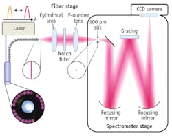
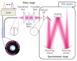
"The principle of our implementation of wavelength-modulated Raman spectroscopy is that fluorescence emission, ambient light, and system transmission function do not significantly vary, whereas the Raman signals do vary upon multiple wavelength excitation with small wavelength shifts," explained corresponding author Christoph Krafft of the Institute of Photonic Technology. "In turn, this leads us to 'cleanly' extract the Raman signature even in the presence of such factors." The researchers' hardware-based approach (see figure) suppresses confounding factors in Raman spectra, requiring minimal pre-processing and offering further advantages.
"This work represents a significant step beyond current Raman microscopy that breaks completely new ground," said Biomedical Spectroscopy and Imaging editor-in-chief Parvez Haris, explaining that biomedical Raman analysis is on the verge of acceptance by the wider community and clinical practice if key issues, such as the ones the authors have raised, can be overcome.
1. S. Dochow et al., Biomed. Spectrosc. and Imaging, 1, 383–389 (2012); doi:10.3233/BSI-120031.

