OPTICAL COHERENCE TOMOGRAPHY/HIGH-SPEED BIOMEDICAL IMAGING: No speed limit: The multi-megahertz approach to optical coherence tomography
A new era has dawned in optical coherence tomography (OCT). Fourier domain mode-locking (FDML) lasers enable sustained imaging rates about 50x that of any other source, with comparable quality. What will high-speed OCT enable, and what impact will it have on the development of future systems?
ByThomas Klein, Wolfgang Wieser, and Robert Huber
Because of its ability to provide three-dimensional (3-D) information on the scattering properties of biological samples, optical coherence tomography (OCT) has become a popular imaging approach in medicine and biology. While the modality has been limited by technological concerns, advances have been steadily improving the applicability of OCT (see sidebar).
Current work around speed improvement opens up a whole new vista for OCT—and implies many benefits for biomedical professionals as well as patients. It also brings up a number of questions for the future.
SS-OCT with Fourier domain mode-locking
Swept-source OCT (SS-OCT) uses a narrowband tunable laser source to enable detection in a mode similar to chirped radar sensing with radio waves. In SS-OCT one OCT depth scan is generated per wavelength sweep—so fast imaging would require sweep repetition at high rates. Unfortunately, standard tunable lasers are inherently limited in their maximum achievable sweep speed by the buildup dynamics of the light field in the laser cavity. Each time the intra-cavity optical bandpass filter, which is required for wavelength tuning, is set to a new wavelength position, lasing has to start from fluorescence background again.19
An approach that completely overcomes this limitation is synchronization of the optical roundtrip time of the light in the laser cavity with the filter sweep time. This technique is called Fourier domain mode-locking (FDML). An FDML laser usable for OCT was demonstrated for the first time in 2005 and initially achieved wavelength sweep repetition rates of 290 kHz.8,20, 21 Buffering has enabled multi-megahertz sweep rates—up to 1.4 MHz for retinal imaging at 1050 nm and up to 5 MHz for imaging in highly scattering tissue.7,22, 17, 18 Sustained OCT imaging rates with FDML lasers of >20 MHz line rate are about 50x faster than any other source with comparable image quality.
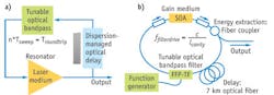
In principle, FDML uses the same cavity components as a standard tunable laser, with an additional very long delay line (see Fig. 1). The delay line usually is a kilometer-long piece of inexpensive standard optical telecom fiber. Light of a certain wavelength is transmitted through the filter, and takes several microseconds (e.g., 1 µs for a fiber with 0.2 km physical length, which is ~0.3 km optical path length) to propagate through the delay fiber. If the filter is tuned (for example, to a repetition rate of 1 MHz), it will be at the same spectral position after 1 µs—which means light of this wavelength can then pass again. Lasing does not have to build up from fluorescence due to feedback from the last wavelength sweep. Thus, FDML represents a real stationary operating regime.23, 24 Consequently, it produces higher output power, improved coherence, reduced intensity noise, and good phase noise performance.8,25, 26
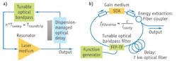
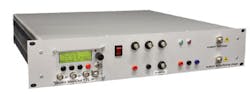
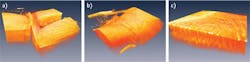
Despite the length of the fiber, such sources can be made very compact: A polarization-maintaining FDML laser source and fiber delay spool can be fully integrated into a 19 in. enclosure (see Fig. 2). FDML is not bound to the type of filter used for wavelength sweeping: Several publications show good performance with polygon mirror-based FDML.27, 28 Because they use telecom components, these sources are inexpensive and highly reliable, and their output performance qualifies them for many biomedical (and non-biomedical) applications.29-42
But, you may ask, “How good is the achievable OCT image quality?” This is a good question because at high speeds and constant power level, exposure increasingly declines, along with the total number of collected photons per depth scan. Considering OCT’s very weak back-reflection signals of -100 dB and less, at some point the signal levels will fall below noise. The incident power on the sample can usually not be increased due to exposure limitations for in-vivo imaging.
OCT imaging in highly scattering tissue
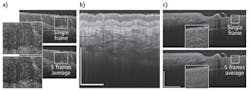
The achievable image quality at multi-megahertz line rates has been investigated in highly scattering tissue using a 1300 nm FDML laser. At line rates up to 5 MHz, good image quality and penetration depth is observed. By applying advanced averaging protocols, the quality can even be further improved (see Fig. 3).
Since the main application of imaging in scattering tissue is intravascular OCT, there have been first trials to improve the speed of today’s commercial OCT systems using FDML lasers: 100 kHz line rates have been achieved so far.43
The ultimate OCT imaging speed can be further pushed by applying a multi-beam scan approach, which has produced a 20.8 MHz A-scan rate with good imaging quality (see Fig. 4).8,25 Such high imaging speeds might be required to investigate transient phenomena in 3-D in real time. The goal is “video rate volumetric imaging.” Video-rate volumetric imaging might become a powerful tool for applications ranging from studies of transient phenomena to real-time surgical monitoring and guidance.
Ophthalmic applications
Today’s biggest market for OCT is ophthalmic imaging. While that area is dominated by retinal imaging, imaging of the eye’s anterior segment is a growing area of interest. Current commercial retinal systems operate at a ~20–50 kHz line rate—meaning only a small amount of data is recorded per acquisition. For this reason, special scan protocols—each of which covers a small subset of the retina—are employed. The most common scan protocols are a star-scan around the macula and a circular scan around the optic nerve head. What this means (to put it bluntly) is that the investigator needs to know what s/he is looking for before scanning, since the scan protocol must be chosen prior to OCT imaging and no corrections can be made afterwards.
Scan rates in the megahertz and multi-megahertz regime promise a significant simplification of the imaging process. Such an advance would mean that a single acquisition could cover a large part of the retina, including both the macula and optic nerve head. Figure 5 shows different projections and visualizations of retinal OCT images at a 1.4 and 6.7 MHz line rate (two beams, 3.4 MHz each), acquired with a 1050 nm FDML laser. The densely sampled 3-D data set provides numerous options for the extraction of information. For example, different “virtual scan paths” can be extracted after the dataset has been acquired. Since all the different scans are derived from a single huge data set consisting of 1900 × 1900 scans, perfect registration is possible.
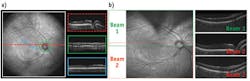
An artificial, high-definition fundus projection, also extracted from the OCT data, can help to exactly locate the different, arbitrarily placeable, linear and circular scans. Moreover, the large field of view covered by the OCT device is for the first time comparable to standard clinical imaging modalities such as fundus photography, enabling a direct comparison and registration. While the acquisition time of 3 s for this dataset may be too long for clinical applications, a further increase in imaging speed may lead to “snapshot” acquisition times of less than a second. This single-shot coverage of the retina will benefit both patient and physician: Reduced acquisition time increases patient comfort and reduces cost, and because substantially more information is collected, diagnostic capabilities are potentially increased.
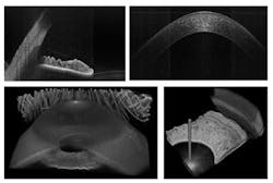
The multi-megahertz FDML also enables the rapid acquisition of 3-D data sets of the entire anterior chamber (see Fig. 6). Because the data set is virtually motion artifact-free, absolute measurements of the geometry are possible.
What’s ahead
Commercial OCT systems running at 30–80 kHz seem destined for eventual replacement by systems running in the several-hundred kilohertz range. Numerous groups and companies are working on alternative approaches of wavelength swept lasers and have already published promising results with wavelength sweep rates up to ~800 kHz. 44-46 Maybe in the future these sources will also achieve megahertz sweep repetition rates.
Whether we’ll ever achieve an increase beyond 1 MHz line rate in commercial systems depends on the answers to three questions:
1) To what extent can we tolerate the image quality reduction that inevitably accompanies higher speeds?
2) How much added value does the speed increase provide for the different applications?
3) What will be the cost of the system?
In turn, the answers to these questions will be dictated by application; it is highly likely that we will see different scan rates for different uses of OCT. So, we can look forward to an exciting future in OCT technology, with the question of the ultimate OCT imaging speed for commercial systems still being open.
Even after 20 years, vital evolution in OCT technology continues to parallel adoption in clinical routine. Ultimately, the patient will reap the rewards of this progress.
REFERENCES
For the list of references, please visit www.bioopticsworld.com/huber/references.html.
Thomas Klein and Wolfgang Wieser are Ph.D. students in the group of Robert Huber in the BioMolecular Optics laboratory at Ludwig-Maximilians University (Munich, Germany); e-mail: [email protected]; www.bmo.physik.uni-muenchen.de.
Improvements to OCT
The slow data acquisition speed of early time domain (TD) OCT systems in the range of ~1 kHz line rate usually limited OCT imaging to single, two-dimensional cross-sections (B-frames, from ultrasound terminology). For instance, at a 1 kHz line rate, it takes 1 s to acquire a B-frame cross-section composed of 1,000 individual depth scans (A-scans). Due to this speed limitation, usually no full 3-D data sets have been acquired with TD-OCT.
The introduction of frequency domain (FD) detection techniques for OCT has led to a dramatic increase in imaging speed.2-6 Line rates of ~50–400 kHz are now common.7-10 At these speeds, it becomes feasible to acquire entire 3-D data sets, offering a greatly improved flexibility in image data visualization, analysis, quantification, and processing.7, 11-13 Further, by using advanced image processing algorithms, 3-D data acquired at high speed can be used to extract functional image information like Doppler flow—so speed can also be used to generate functional image contrast.14
Even though all these applications already demonstrated the tremendous potential of 3-D OCT imaging, in most cases the 3-D data sets had highly unbalanced numbers of samples in each of the three dimensions, or the acquisition time was too long for routine in-vivo imaging of non-trained patients. Current ophthalmic OCT systems either cover only a fraction of the eye background or they apply sparse sampling; i.e., in one direction the spacing of the individual OCT A-scans is much larger than the transverse optical resolution. In endoscopic applications for intravascular imaging, a typical spiral sampling pattern has a spacing of >100 µm at transverse optical resolutions of 30 µm, also resulting in sparse sampling. Here, the maximum time during which the blood can be cleared by flushing with a contrast agent limits the total number of A-scans.
For many applications, the area of interest can be densely covered by several million OCT-scans.
Imaging time for in-vivo OCT applications is often limited to a few seconds, making scan rates in excess of 1 MHz highly desirable. However, the main problem impeding the increase of imaging rate is the requirement to maintain good image quality. Some early publications with megahertz line rates exhibited only limited image quality.15, 16 Recently, OCT systems using a so-called Fourier domain mode-locked (FDML) laser demonstrated that good image quality can be achieved with OCT at line rates beyond 1 MHz.8, 17, 18
More BioOptics World Current Issue Articles
More BioOptics World Archives Issue Articles