Components Systems & Products
Compiled By LEE MATHER
Please send your new product announcement to [email protected]
Quantitative fluorescence imaging light sources
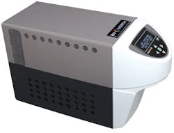
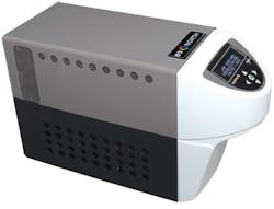
The PhotoFluor II and PhotoFluor II NIR light sources for quantitative fluorescence imaging pair a self-aligning, 200 W metal-halide lamp with a sputtered, infrared (IR)-blocking filter to enable transmission in the UV and NIR, respectively. PhotoFluor II works in the 340-650 nm wavelength range, while PhotoFluor II NIR works from 360 to 800 nm. Both accommodate imaging of several fluorescence probes, including-but not limited to-green fluorescent protein (GFP), mCherry, and Texas Red.
89 North, Burlington, VT, http://bit.ly/GFKheQ
Dual-view imaging system
A dual-view imaging system enables simultaneous view of a single subject and image capture at multiple fields of view (FOV) with a single objective lens presented to two separate imaging channels. The channels can be fixed, zoom, or a combination of both. Two separate light sources and wavelengths can be used, allowing inspection of multiple types of materials. Maximum optical magnification using the system can range from 20 to 583x using a 50x objective.
Navitar, Rochester, NY, http://bit.ly/H7JeVC
Retinal hyperspectral imaging system
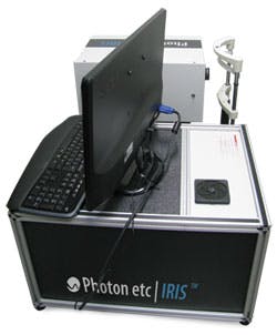
The IRIS retinal hyperspectral imaging system delivers noninvasive localization of biomolecules in the eye fundus to develop treatments for retinal diseases such as diabetic retinopathy and age-related macular degeneration (AMD). Based on a mydriatic retinal camera and tunable laser source, the system allows high-definition retinal imaging from 420 to 1000 nm. A spectral calibration setup enables spectral reproducibility, while an integrated photodiode provides temporal normalization of light intensity.
Photon etc., Montreal, QC, Canada, http://bit.ly/xDCFSn
Light-based melanin reader
The Skintel Melanin Reader determines average melanin density in an area of skin to be treated, in a quantitative manner, prior to a light-based aesthetic treatment. The reader measures skin diffuse reflectance at three wavelengths of light and computes these values into a Melanin Index value, which wirelessly sets the suggested parameters in the company's Icon Aesthetic System for a starting treatment test spot fluence.
Palomar Medical Technologies, Burlington, MA, http://bit.ly/HQAW2r
Laser objective
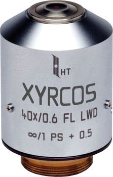
The XYRCOS laser objective for advanced research applications integrates a laser and the company's RED-i target locator, and works with all major microscope models. An enhanced UV transmission/fluorescence is compatible with many fluorescing stains used in applications such as creating transgenic animals, gene targeting, and stem cell research. Both 40X and 20X versions are available, with the company's Staccato multi-pulse laser activation software as an option.
Hamilton Thorne, Beverly, MA, http://bit.ly/GAY0DG
11 Mpixel camera for scientific imaging
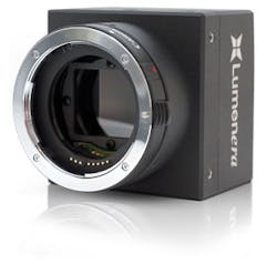
The Lg11059 11 Mpixel Gigabit Ethernet (GigE) digital camera delivers 5 fps at full 4008 × 2672 resolution for scientific imaging applications with low-light conditions, such as in pathology. The camera's CMOS image sensor with a 43.3 mm image diagonal and electronic global shutter delivers low dark current noise, negligible lag, and low smear.
Lumenera, Ottawa, ON, Canada, http://bit.ly/I3t01a
Balanced receiver for OCT
The EBR370000 balanced receivers cover wavelengths from 400 to 1600 nm to support applications like optical coherence tomography (OCT), which requires high signal-to-noise performance. The receivers' bandwidth is continuously adjustable from 60 to 380 MHz. Features include a high common-mode rejection ratio, electronically switchable gain, two monitor outputs (DC-400 kHz), and a 5 V power supply.
Exalos, Zürich, Switzerland, http://bit.ly/HTwK2r
Image analysis software
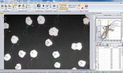
Image-Pro Premier image processing and analysis software provides tools to capture, process, enhance, measure, compare, analyze, automate, and share images and valuable data for applications such as brightfield microscopy, cell biology, fluorescence imaging, and marine biology. The software can stream multi-gigabyte movies directly to the host computer's hard drive and it supports multiresolution files, including Aperio (.svs) and BigTIFF (.btf).
Media Cybernetics, Bethesda, MD, http://bit.ly/IzhRmY
More BioOptics World Current Issue Articles
More BioOptics World Archives Issue Articles
