FLUORESCENCE MICROSCOPY/IMAGING PROBES: Super-efficient fluorophor will facilitate cell research
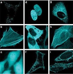
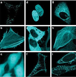
A molecule that emits turquoise light more efficiently than anything seen before in living cells promises to improve cell imaging, and enable the study of protein-protein interactions in living cells with unprecedented sensitivity.1
The probe was developed by a team led by Antoine Royant at the Institut de Biologie Structurale/CNRS-CEA-Université Joseph Fourier (Grenoble, France). Using highly brilliant X-ray beams at the European Synchrotron Radiation Facility (ESRF; Grenoble, France), scientists from Grenoble and from the University of Oxford in England discovered details of how cyan fluorescent proteins (CFPs) store incoming energy and retransmit it as light: They resolved the molecular structures of different CFP crystals and learned that a subtle process near the chromophore could be manipulated to improve fluorescence efficiency. "We could understand the function of individual atoms within CFPs and pinpoint the part of the molecule that needed to be modified to increase the fluorescence yield," explained David von Stetten, ESRF. In parallel to this work, a team at the University of Amsterdam (The Netherlands), led by Theodorus Gadella, used an innovative screening technique to study hundreds of modified CFP molecules, measuring their fluorescence lifetimes under the microscope to identify which had improved properties. The result of the combined structural and cellular biology efforts is mTurquoise2, a CFP with a fluorescence efficiency of 93%. The scientists now hope to improve fluorescent proteins of other colors, too.
1. J. Goedhart, Nat. Comm., 3, 751 (2012).
More BioOptics World Current Issue Articles
More BioOptics World Archives Issue Articles

