MICROSCOPY/CELL BIOLOGY: Label-free 3D technique visualizes sub-cellular noninvasively
A fast, noninvasive 3D microscopy approach has proven able to visualize, quantify, and study cells without the use of fluorescence or contrast agents, according to scientists at the University of Illinois at Urbana-Champaign (Champaign, IL).
The technique is based on spatial light interference microscopy (SLIM), which was designed by Beckman Institute researcher Gabriel Popescu as an add-on module to a commercial phase contrast microscope. SLIM is extremely fast and sensitive at multiple scales (from 200 nm and up), but its resolution is limited by diffraction.
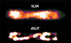
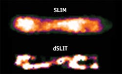
By applying a novel deconvolution algorithm to retrieve sub-diffraction limited resolution information from the fields measured by SLIM, Popescu and fellow researchers were able to render tomographic images with a resolution beyond SLIM's limits. They used the sparse reconstruction method to render 3D reconstructed images of E. coli cells at sub-cellular resolution.
Last year, the same research group demonstrated a new optical technique called spatial light interference tomography (SLIT) that enables 3D measurements of complex fields on live neurons and photonic crystal structures. For this project, they developed a novel algorithm to further extend the 3D capabilities by performing deconvolution on the measured 3D field, based on modeling the image using sparsity principles. This microscopy capability, called dSLIT, was used to visualize coiled sub-cellular structures in E. coli cells.
The researchers note that the new method addresses two major issues in cell microscopy: Lack of contrast, due to the thin and optically transparent nature of cells, and diffraction-limited resolution. They note that dSLIT's ability to retrieve limited resolution information delivered by SLIM will give researchers a tool to study structures in a new way—and thus may enable new insights.
1. M. Mir et al., PLoS ONE, 7, 6, e39816 (2012).

