MICROSCOPY/CELL BIOLOGY: TIRF and confocal microscopy illustrate wound healing dynamics
Cell migration is critical to such biological functions as wound healing and cancer metastasis, which require that many cells move together. Now, total internal reflection fluorescence (TIRF) microscopy is helping to understand how endocytosis—the process by which cells take in substances from their surroundings—could play a key role in the cell migration required for wound repair to the skin and internal organs.1
Working in the Birmingham Advanced Light Microscopy (BALM) facility, researchers at the University of Birmingham (England) have demonstrated that migrating cells take in proteins from their surface. To learn which of these proteins was the most important, the team identified two candidate molecules that they suspected played a key role in migration: These proteins help to anchor a cell in place and help it detect environmental changes.

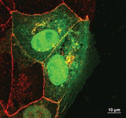
The team initially expected that the cells on the edge of a wound would use endocytosis to draw in the molecules that make structures called focal adhesions, which allow a cell to cling to its surroundings. However, TIRF revealed that endocytosis was not involved in rearranging the focal adhesions during wound healing. Confocal microscopy then showed that a different protein, occludin, was being taken in via endocytosis.
In healthy tissue, occludin holds epithelial cells together to form a single flat layer. When the researchers created a layer of epithelial cells in culture and then made a wound, the cells on the edge pulled occludin in via endocytosis and then moved forward to fill the gap. But when the researchers added additional occludin to the spaces between cells, the wound was not filled as quickly.
1. S. J. Fletcher, N. S. Poulter, E. J. Haining, and J. Z. Rappoport, Biol. Cell, doi: 10.1111/boc.201100004 (2012).
More BioOptics World Current Issue Articles
More BioOptics World Archives Issue Articles