OPTICAL MICROSCOPY/OPTOGENETICS: Simultaneous, multi-color scanning with diode lasers
Fifty years since the advent of laser diodes, technology advances including beam manipulation and multi-laser engines heighten the impact of this technology on life sciences.
ByMarion Lang
Lasers have played a critical role in the growth of biomedicine and while gas lasers have dominated the field for more than two decades, they are now being replaced by more compact and cost-effective laser types, including diode lasers.
Diode lasers and laser diodes
This year, 2012, the laser diode celebrates its 50th anniversary: In 1962, lasing was observed in semiconductor diodes for the first time. This discovery enabled economical mass production of lasers small and inexpensive enough to make them practical for applications that other lasers had been unable to reach, with the result being the launch of the Internet, among other paradigm-shifting technological developments. Today, laser diodes are used in CD and DVD players (780 and 655 nm, respectively), telecom networks (1550 nm), and Blu-ray players (405 nm)—in addition to biomedicine. In fact, biophotonics has benefitted greatly from these mass-market developments: Laser diodes are compact, convenient, cost-effective, and highly efficient. However, standard laser diodes have properties—such as poor beam profile and imprecise lasing wavelength—that are undesirable for certain applications.
A diode laser—that is, a whole system including optics, electronics, and a laser diode semiconducting element—provides additional capabilities that ensure that the output of the device has the right properties. For example, atom optics experiments require a laser with a very narrow linewidth, and the emission of a plain laser diode is much too broad—but integrated gratings can enable precise selection and application of the appropriate wavelength. And for microscopy, a TEM00 beam profile is highly desirable—but the beam originating from a laser diode is highly elliptical, so special optics are required to transform this to a Gaussian profile.
Compact and efficient, diode lasers are a welcome replacement for bulky gas lasers. As of 2004, annual unit sales of diode lasers was approximately 733 million,1 while about 131,000 of other laser types were sold in the same time period.2 A significant advantage compared to gas and diode-pumped solid-state (DPSS) lasers is that diode lasers can be directly modulated with up to 250 MHz and require no acousto-optical modulator (AOM).
Multi-laser engines
Let's take a look at diode lasers and their integration into multi-laser engines, which combine two or more lasers into a single housing and thus provide several wavelengths to excite different dyes in the sample. We'll discuss how these technologies have enabled multi-color experiments, which are relevant for colocalization and essential for understanding the spatial organization of different structures with respect to one another (see "State-of-the-art diode lasers and multi-laser engines" for a discussion of current product offerings).
While a number of tunable light sources are able to facilitate multi-color experiments, such solutions tend to be expensive and complex (in the case of a combined Ti:sapphire laser and an optical parametric oscillator [OPO]) or else insufficiently powerful (in the case of a supercontinuum source [a.k.a. "white light laser"] generating 460 nm to 2 μm, but using a photonic crystal fiber [PCF] or a source that provides a narrow-linewidth output that is tunable from 488 to 640 nm [e.g., Toptica's iChrome TVIS]). Still, however, 405 nm is one of the wavelengths that can only be generated with gas or diode lasers.
The wavelength of a diode laser depends on the bandgap of the semiconductor material used: Laser diodes based on gallium arsenide (GaAs) and indium phosphide (InP) cover a wide wavelength range (especially blue and red wavelengths) and only recently, green laser diodes emitting at 515 nm became available. Laser diodes based on GaAs material offer wavelengths above 630 nm, including 640, 660, 685 and 785 nm. Laser diodes based on InP offer many shades of blue; for instance, 375, 395, 405, 420, 445, 460, 473, 488, and 515 nm. Because an increasing number of applications require more than one excitation wavelength, lasers with different wavelengths are being combined in instruments.
Microscopy methods
Compared to a point-scanning confocal microscope, fast imaging techniques for live-cell microscopy generally require the higher output powers that diode lasers and multi-laser engines provide.
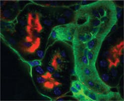
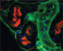
One such technique, spinning disk confocal microscopy, parallelizes the imaging process and instead of scanning with a single beam, uses multiple beams to scan the specimen (see Fig. 1). This parallel approach provides higher frame rates compared to confocal microscopy, which makes these techniques especially useful for live-cell imaging.
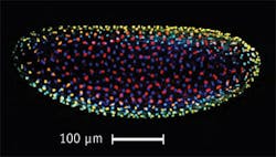
Another fast imaging method, known by light-sheet fluorescence microscopy (LSFM) and selective plane illumination microscopy (SPIM), allows examination of processes in live, fluorescently labeled specimens both large (animals) and small (cells)—even in deep tissue layers. Here, a sheet of light illuminates the sample and a charge-coupled device (CCD) camera in an orthogonal plane detects the fluorescence light. The technique achieves an optical sectioning effect similar to confocal microscopy, but with bleaching limited to the imaged plane only. Since the complete plane is imaged all at once, this technique also acquires images much faster than conventional confocal microscopy (see Fig. 2).
Total internal reflection fluorescence (TIRF) microscopy is well suited for the analysis of cellular processes near the plasma membrane. Specialized objectives illuminate the sample at the so-called "critical angle." At the interface between the glass cover slip and the aqueous sample, total internal reflection takes place. An evanescent field extends into sample approximately 100 nm and excites the fluorophores within this area. This technique minimizes background noise introduced by cytoplasm, for instance, and in this way achieves a higher contrast compared to other techniques. Multicolor excitation allows discernment of components such as actin, prestin, and a cell nucleus.
Enabling optogenetics
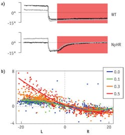
Optogenetics combines optics and genetics to control the activity of cell groups in organisms. The technique depends upon opsins, a class of light-sensitive receptors that includes channelrhodopsin, an ion-channel that can be activated with blue light, and halorhodopsin, an ion-pump that can be switched on with red light. Excitable cells coexpressing these proteins can be activated/inhibited with blue/red light pulses, respectively. Controlling the activity of the cells requires a precise spatial and temporal stimulation of the structures with light pulses. Aristides Arrenberg of the University of Freiburg (Germany) activates these light-sensitive proteins in zebrafish larvae by positioning the fiber output of his multi-laser engine above the head of the transparent animal. By activating and inactivating groups of neurons expressing the proteins, Arrenberg controls neural circuits and tests their function in a behaving animal (see Fig. 3).
Flexible, compact, and easy-to-use multi-laser engines enable other biological applications as well, including flow cytometry. In fact, diode lasers have enabled the design of convenient, tabletop flow cytometry systems. Because their integration and operation is very straightforward, the lasers can take their part in complex biological experiments, without need for special attention.
REFERENCES
1. R.V. Steele, "Diode-laser market grows at a slower rate," Laser Focus World, 41, 2 (2005).
2. K. Kincade and S. Anderson, "Laser Marketplace 2005: Consumer applications boost laser sales 10%," Laser Focus World, 41, 1 (2005).
Marion Lang, Ph.D., is Technical Marketing Manager for Toptica Photonics AG (Gräfelfing, Germany), www.toptica.com. Contact her at [email protected].
State-of-the-art diode lasers and multi-laser engines
Recent developments in diode lasers include the provision of more colors: From 375 nm to 515 nm and from 640 nm to infrared (IR), diode lasers can generate virtually any wavelength. The new generation of technology also provides increased output powers, such as 300 mW at 405 nm and 200 mW at 488 nm, all from single-mode laser diodes. The lifetimes of diodes have also increased over the last years—now, lifetimes of more than 100,000 hours can be achieved, even in UV.
Until recently, multi-laser engines used "breadboard approaches"—that is, several lasers were mounted on a breadboard and combined with dichroic mirrors, and finally coupled into an instrument such as a microscope. These approaches can be affected by ambient conditions, however, so long-term power stability may be a problem. And such systems must be realigned by skilled persons from time to time, to adjust the beams for high output powers. The new generation of multi-laser engines overcomes these issues.
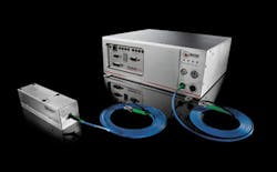
An example of a tool that addresses the increasing use of multiple colors in experiments is the iChrome MLE, a multi-laser engine that houses up to four diode lasers—or three diode lasers and one DPSS laser—and delivers all wavelengths via one optical fiber (see figure). Different wavelength combinations allow adaption of the system to specific requirements of an experiment. All lasers can be switched on and off completely independently with up to 20 MHz, which allows for arbitrary excitation patterns. A single-mode, polarization-maintaining fiber delivers up to 100 mW per wavelength to the microscope.
The iChrome is offered with COOLAC (Constant Optical Output Level) technology, which will benefit everyone who has ever tried to align several laser sources. Such a setup requires occasional fine-tuning due to thermal drift—a task that is especially challenging when coupling lasers into a single-mode fiber with a core diameter of 4–6 μm. COOLAC allows fully automatic push-button alignment of the lasers and thus ensures permanently highest output powers after fiber delivery.
