STIMULATED RAMAN SCATTERING/LIVE CELL IMAGING: Chemical tag and SRS combo images small biomolecule dynamics within living cells
The ability to visualize small biomolecules inside living biological systems with minimal disturbance has been a goal in the scientific community for years. When studying intracellular function, researchers typically label molecules of interest with fluorophores. With small biomolecules, though, such tagging is problematic: A fluorophore the same size or larger than the molecule of interest disturbs the function of molecules with crucial biological roles.
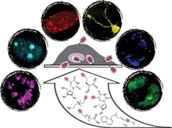
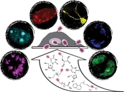
Led by Wei Min of Columbia University (New York, NY), a team of scientists from Columbia and Baylor College of Medicine (Houston, TX) has developed a general method to determine the exact location and function of targets such as small biomolecule drugs, nucleic and amino acids, and lipids.1 The approach "could do for small biomolecules what fluorescence imaging of fluorophores such as green fluorescent protein (GFP) has done for larger species," said Min. Instead of fluorescence, though, the researchers turned to a novel combination of physics and chemistry: They coupled an emerging laser-based technique called stimulated Raman scattering (SRS) microscopy with a tiny, highly vibrant alkyne tag—a carbon-carbon triple bond that, when stretched, produces a strong Raman scattering signal that is distinct from natural molecules.
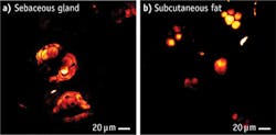
Alkyne labeling avoids the perturbation inherent in fluorescence tagging while leveraging the extreme specificity and sensitivity of SRS. By tuning the laser colors to the alkyne frequency and quickly scanning the beam across the sample, point-by-point, SRS microscopy detects the stretching motion of the C=C bond and produces a three-dimensional map of molecules within living cells and animals. In this way, Min's team tracked alkyne-bearing drugs in mouse tissues, and visualized de novo synthesis of DNA, RNA, proteins, phospholipids, and triglycerides through metabolic incorporation of the alkyne-tagged precursors in living cells.
"The major advantages of our technique lie in the superb sensitivity, specificity, and biocompatibility with dynamics of live cells and animals for small molecule imaging," said lead author and Columbia Ph.D. candidate Lu Wei.
Next, the team will apply its technique to such applications as detecting tumor cells and probing drug pharmacokinetics in animal models. The researchers are also creating other alkyne-labeled biologically active molecules for more versatile imaging applications. The approach will "open up numerous otherwise difficult studies on small biomolecules in live cells and animals," said Min. "In addition to basic research, our technique could also contribute greatly to translational applications."
1. L. Wei et al., Nat. Meth., 10, 12, 1177–1184 (Dec. 2013); doi:10.1038/nmeth.2878.


