MICROSCOPY/LIVE CELL IMAGING/CELL BIOLOGY: First STED imaging of live cells in two colors

Sept. 1, 2011
Stimulated emission depletion (STED) microscopy—a super-resolution approach that reveals cells and cellular components in detail by absorbing and releasing energy in fluorescent dye—has been limited to single-color imaging of living cells.

Stimulated emission depletion (STED) microscopy—a super-resolution approach that reveals cells and cellular components in detail by absorbing and releasing energy in fluorescent dye—has been limited to single-color imaging of living cells. But effective study of active cell processes, such as protein interactions, really requires multicolor imaging. Now, a team of researchers from Yale University has given a boost to cell biology by reaching this goal. The group describes its work in the August 2011 issue of Optics Express.1

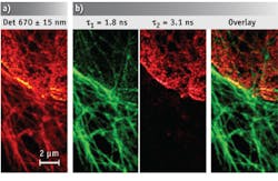
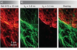
Tubulin and lamin, immunostained with ATTO 647N and KK 114, respectively; a) Raw intensity STED data, and b) channels decomposed by lifetime separation (green: tubulin, red: lamin). (Images courtesy Optics Express)

The key to the researchers’ achievement was overcoming the challenges of labeling target proteins in living cells with dyes optimal for two-color STED microscopy. By incorporating fusion proteins, they improved the targeting between the protein and the dye, effectively bridging the gap. This enabled them to reach resolutions of 78 and 82 nm for 22 sequential two-color scans of two proteins—epidermal growth factor and epidermal growth factor receptor—in living cells.

The researchers expect that this and other novel approaches will expand live cell STED microscopy to three and more colors, eventually enabling imaging in three dimensions.

1. J. Bückers et al., Opt. Exp. 19, 3130–3143 (2011).

More BioOptics World Current Issue Articles
More BioOptics World Archives Issue Articles
Sponsored Recommendations

Optical Power Meters for Diverse Applications

April 30, 2024
Bench-top single channel to multichannel power meters, Santec has the power measurement platforms to meet your requirements.

Request a quote: Micro 3D Printed Part or microArch micro-precision 3D printers

April 11, 2024
See the results for yourself! We'll print a benchmark part so that you can assess our quality. Just send us your file and we'll get to work.

Request a Micro 3D Printed Benchmark Part: Send us your file.

April 11, 2024
See the results for yourself! We'll print a benchmark part so that you can assess our quality. Just send us your file and we'll get to work.

Request a free Micro 3D Printed sample part

April 11, 2024
The best way to understand the part quality we can achieve is by seeing it first-hand. Request a free 3D printed high-precision sample part.

Voice your opinion!

To join the conversation, and become an exclusive member of Laser Focus World, create an account today!