BIOINSTRUMENTATION/NEUROSCIENCE: Flexible, inexpensive mini microscope enables in-vivo discovery
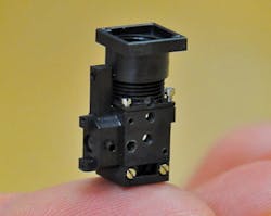
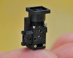
A fluorescence microscope that weighs just 1.9 g and incorporates all optical components (LED, image sensor, filters, and microlenses) in a 2.4 cm3 housing has demonstrated advantages over previous approaches and facilitated neurology discoveries in living mice. Because all components of the miniature microscope are batch-fabricated, the unit is economical. And because it doesn’t require optical realignment when transported, it is easily portable.1
Compared with fiberoptic technology, the unit offers a ~700% greater field of view and ~500% greater transfer efficiency of fluorescence between the specimen and image detection planes, the researchers claim. It attaches by floppy tether to the head of a freely moving adult mouse; the fine electrical wires—which carry power, control signals, and data—allow flexibility instead of exerting torque as optical fibers do.
Researchers at Stanford University (Stanford, CA) developed the device and tested it in a range of applications, including the visualization of cerebellar microcirculation in active mice, wherein it demonstrated sensitivity and image quality sufficient to track erythrocyte speeds and capillary diameters with two-second time resolution. Motion artifacts were typically <1 µm, even during running—substantially less than during two-photon microscopy in head-fixed behaving mice, the team reports. Contrary to the researchers’ expectations, their work indicates that capillaries separated by only tens of micrometers are controlled nonuniformly, and that a subset of vessels appears to dominate bulk effects.
In what may be the first analysis of higher-order correlations in cell dynamics in freely behaving mice, the device also enabled the team to track Ca2+ spiking in more than 200 individual cerebellar Purkinje neurons at 46 Hz—compared with the ability of two-photon microscopy, which has enabled tracking of about 100 Purkinje neurons at less than 12 Hz.
The researchers also demonstrated the device’s capabilities in fluorescent cell counting, and detecting tuberculosis bacteria in fluorescence assays. Further, they suggest usefulness in microscope arrays for high-throughput screens (current approaches do not involve imaging), imaging within instruments such as incubators, or combinations with other integrated components such as microfluidic or gene chips.
1. K.K. Ghosh et al., Nat. Meth. 8, 871–878 (2011).
More BioOptics World Current Issue Articles
More BioOptics World Archives Issue Articles

