Bioinspired imaging system takes notes from shrimp
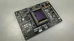
Inspired by the multiwavelength capabilities of a mantis shrimp’s vision, a team of researchers at the University of Illinois at Urbana-Champaign (UIUC) has developed a compact camera on a single chip. It can capture images in the ultraviolet, near-infrared, and visible ranges, much like the marine crustacean’s vision, which separates different wavelengths of light in a compact space (see video).
“Our goal was to build a compact surgical camera that can see more than a standard color image,” says Viktor Gruev, a professor at UIUC. Targeting lymph nodes, an important part of the lymphatic system, “our imager captures visible light, near-infrared (NIR) fluorescence, and ultraviolet (UV) autofluorescence from the same field of view.”
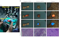
Each wavelength reveals something different: The visible image gives the surgeon a familiar view of the tissue, while the NIR signal that uses indocyanine green (ICG) helps locate the lymph nodes that drain from the tumor area. And the UV signal provides a label-free way to look at the tissue itself and detect optical changes that may be associated with cancer.
“We used a mantis shrimp biological model as inspiration for a single-chip sensor with pixel-level filters and stacked photodiodes, which allows the camera to collect that UV, visible, and NIR information—without needing multiple cameras,” Gruev says.
The single-chip imager offers real-time information on lymph nodes’ location via NIR imaging. It also uses UV imaging to determine whether they appear suspicious for cancer.
Pros and cons
The new system combines two important functions: Finding the lymph node and helping assess whether it may contain cancer.
ICG fluorescence can help surgeons find lymph nodes, but it does not tell whether those nodes are malignant. Pathology can answer that question, but often not in real time during surgery. Some label-free optical methods can provide the biochemical contrast needed to detect cancer, Gruev explains, but those are not typically combined with other imaging modalities.
“To provide both the location and likely cancer status of a lymph node requires acquiring images using different wavelengths of light while keeping those images carefully aligned,” he says. “Our approach is designed to help fill the gap.”
The signals are collected on the same sensor, so the UV, visible, and NIR images remain aligned. Gruev notes this is important in surgery, where tissue and instruments are moving and the information needs to be easy to interpret, and quickly.
Findings of note
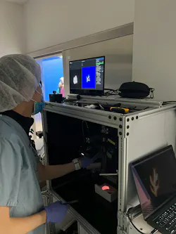
The team tested their system in stages in an ex vivo breast cancer study of freshly removed tissue from 33 patients. During controlled lab tests, the camera was able to capture UV, color, and NIR information together. It also measured its sensitivity and accuracy. Analyzing 94 sentinel lymph nodes, they found metastatic lymph nodes produce a stronger UV autofluorescence signal than nonmetastatic nodes.
The NIR channel helped locate the nodes, while the UV channel helped distinguish metastatic from benign nodes. In the independent validation set, the UV signal achieved an AUC (area under the curve) of 0.89, with 97% sensitivity and 89% specificity.
“For me, the exciting point is that this was done with a compact imaging platform that combines localization and tissue assessment in one system,” Gruev says.
What’s next?
The team now plans to shift from an ex vivo research system to a practical intraoperative tool.
“It means making the system faster, easier to use, and fully compatible with the operating room,” Gruev says.
They will also need to conduct larger clinical studies to test the approach across more patients and more clinical settings. Another important step will be to improve the UV sensitivity to produce stronger signals with shorter exposure times and lower light levels. Any clinical instrument using UV light will need careful dose control, as well, and built-in safety monitoring.
“Our ultimate goal is to give surgeons useful optical information in real time, without disrupting the normal surgical workflow,” Gruev says.
Broader goals
True positive outcomes in cancer treatment will require more precise surgery.
“Cancer surgeons are always balancing two priorities: Removing enough tissue to treat the disease, while preserving as much healthy tissue as possible,” Gruev says. “For lymph node surgery, that balance is especially important.”
Removing too many lymph nodes can increase complications such as lymphedema, the swelling of tissue caused by an accumulation of protein-rich fluid. This fluid is usually drained through a healthy lymphatic system.
Missing the lymph nodes involved can affect staging and treatment. “A tool like this could help surgeons make more informed decisions during the operation,” Gruev says.
Beyond this, surgical cameras could potentially do more than just show anatomy. They may also provide real-time optical information about tissue biology, which could ultimately help move cancer surgery toward more selective, personalized, and confident decision-making.
FURTHER READING
Y. Jin et al., Optica, 13, 4, 800-809 (2026); https://doi.org/10.1364/optica.582293.
Related Article
About the Author
 Justine Murphy
Justine Murphy
Multimedia Director, Digital Infrastructure
Justine Murphy is the multimedia director for Endeavor Business Media's Digital Infrastructure Group. She is a multiple award-winning writer and editor with more 20 years of experience in newspaper publishing as well as public relations, marketing, and communications. For nearly 10 years, she has covered all facets of the optics and photonics industry as an editor, writer, web news anchor, and podcast host for an internationally reaching magazine publishing company. Her work has earned accolades from the New England Press Association as well as the SIIA/Jesse H. Neal Awards. She received a B.A. from the Massachusetts College of Liberal Arts.