Modular designs advance Raman spectrometers
Ian R. Lewis and Kevin L. Davis
Interchangeable components offer high performance and versatility in spectrometers that can be easily upgraded for spectroscopic research.During the past several years, Raman spectroscopy has been transformed into a powerful, real-time analytical tool for both research scientists and process engineers. It is used in a wide range of chemical, pharmaceutical, and biomedical applications. Specifically configured integrated instruments have been developed for analytical, monitoring, and online processes. Designers of integrated spectrometers select components based on criteria such as size, cooling needs, and cost. These design criteria, however, may not address issues such as very high resolution, versatility, and ability to upgrade. In response, "plug and play" modular spectrometers, in which fiber-coupled modules can be easily interchanged without optical alignment, have been developed.
Raman background
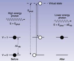
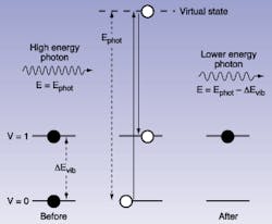
When a high-energy photon interacts with a molecule, there is a small probability of a Raman scattering event occurring (see Fig. 1). The Stokes-shifted photon is essentially "re-emitted" instantaneously at a lower energy, where the photon energy shift (nexcitation - nRaman) is identical to the vibrational frequency of the chemical bond. Consequently, when a monochromatic light source is used to excite a sample, dispersion of the Raman scattered light reveals a vibrational spectrum. Such a spectrum can be used for qualitative and quantitative sample analysis, since each fundamental vibrational band originates from a chemical bond.
Raman spectroscopy combines the advantages of several other spectroscopic techniques. It produces sharp, well-resolved spectra, and relative concentration information often can be derived from measurement of spectral peak intensities. Raman often requires no sample extraction or preparation, and is well-suited to direct observation of aqueous solution samples because water produces only a weak Raman signal. Raman can be carried out at visible and near-infrared (NIR) wavlengths, allowing remote sensing via fiberoptics or in conjunction with optical microscopes for high-resolution chemical Raman spectroscopy. In addition, spectrometers can be designed around high-quality Raman spectroscopy optics and charge-coupled device (CCD) detectors.
Nonetheless, Raman was rarely used for analytical work before the 1990s because of the practical issues of recording the spectra. The main problem was that the Raman scattering effect is very weak, involving as few as 10-7 photons. Traditionally, acquiring a Raman spectrum required the use of complex, costly equipment, often covering an entire tabletop. This equipment required constant expert alignment and maintenance.
The growth of several critical technologies have now made compact, turnkey spectrometers possible. The key technologies enabling this change are small, stable lasers for efficient reliable sample excitation; holographic gratings and holographic notch filters to eliminate Rayleigh scattered laser light and efficiently disperse the Raman spectrum; and cooled CCD detectors that allow simultaneous detection of the entire dispersed spectrum. Rugged, reliable instruments based on these advances have allowed rapid expansion into Raman applications from biomedical Raman spectroscopy to real-time monitoring of pharmaceutical crystallization, and industrial polymer fibers and yarns.
Modular solution
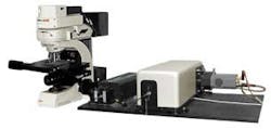
A fully integrated Raman spectrometer clearly is best suited for analytical, monitoring, or process analysis applications. But, in a research environment, instrument versatility, and the ability to upgrade quickly are just as important as turnkey operation. Fortunately, the modular generation of Raman spectrometers delivers all these advantages, making use of easily removable and interchangeable open-architecture modules with efficient fiberoptic coupling (see Fig. 2).
The principal components in a Raman spectrometer are the laser, the holographic notch filters, a Raman spectroscopy spectrograph with a holographic grating, the CCD detector, the sampling accessory/interface, and data acquisition/analysis software. The latest modular spectrometers provide simple, interchangeable options for all these key components without compromising system performance, and without requiring any system realignment. To understand the need for this versatility, it is useful to examine the role of each modular component.
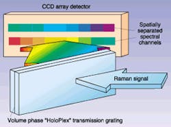
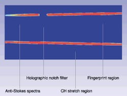
FIGURE 4. HoloPlex holographic, volume-phase, dual-transmission gratings act as two diffraction gratings, separately dispersing the short- and long-wavelength components on the CCD detector. Operation of is shown (top) with actual CCD image (bottom) .
Laser source
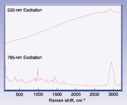
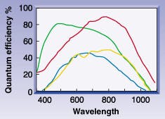
The main factors that influence the choice of laser wavelength are the strength of the Raman signal and the amount of fluorescence excited in the sample. The intensity of the Raman signal is proportional to 1/l4. Fluorescence, however, is more likely at visible wavelengths, as the photon energy reaches excited states. Because fluorescence is many orders of magnitude more efficient than Raman scattering, even a small amount of sample fluorescence can completely swamp and obscure the Raman signal. For many real-world samples, moving to NIR-excitation wavelengths reduces the probability of fluorescence. As a result, shorter wavelengths such as 514.5 and 532 nm are used for known "nonfluorescing" samples such as inorganic pigments, select thin films, and catalysts, whereas longer wavelengths, such as 785 and 830 nm, are generally preferred for unknowns when fluorescence may be encountered, such as petrochemicals, pharmaceuticals, polymers, and biomaterials. The 785-nm diode laser is often favored because at longer wavelengths, silicon CCDs cannot efficiently detect the entire Raman spectrum (see Fig. 3; Laser Focus World, September 2000, p.161).
Spectrograph and gratings
Because the Raman effect is weak, it is vitally important to maximize optical efficiency throughout the spectrometer. Of course, the heart of the spectrometer is the spectrograph itselfwhere the incoming light from the sample is dispersed into its component wavelengths. The use of high-performance transmission holographic gratings allows these modular spectrometers to be designed for f number values as low as 1.4. Also, by designing the spectrograph for efficient fiberoptic input, the optical efficiency is high no matter what type of sampling probe is attached to the system.
The design and choice of diffraction gratings are also critical. The resolution and spectral coverage of the instrument is determined by the dispersion of the grating, the slit width, and the size and number of pixels in the CCD array detector. In many types of optical spectrographs, there is an inherent trade-off between resolution and spectral range. Kaiser Optical Systems developed the HoloPlex grating to address this limitation. This holographic element produces twice the normal spectral coverage, at any given resolution, because the long and short wavelengths are separately dispersed along the long axis of the CCD detector (see Fig. 4). For example, up to 4400 cm-1 can be simultaneously detected at 5-cm-1 resolution with 532-nm excitation. This approach significantly reduces data acquisition time for qualitative measurements and can yield quantitative information, even for chemically changing samples or species with widely separated analytical bands of interest.
The modular approach allows end users to select a grating in which peak transmission is matched to the desired laser excitation wavelength, to deliver maximum signal to noise. Optional high-dispersion and low-dispersion gratings allow the end user to maximize the resolution/range trade-off for a specific type of application.
CCD detector
Selection of an optimal CCD camera is perhaps the most important option in a versatile Raman spectrometer. Considerations include the size and number of pixels, the quantum efficiency curve, and the mode of cooling. The most commonly used CCD formats are 1024 x 128 pixels and 1024 x 256 pixels. The preferred arrangement is normally 128 vertical pixels. But in some applications, multiple fiberoptic inputs from different sampling ports can be stacked vertically at the spectrograph input. In this case, 256 vertical pixel format is the better choice. For very high-resolution spectroscopy, another useful format is a 2048 x 512 chip.
For the researcher, the choice of back-illuminated vs. front-illuminated CCD detectors can be very important. Back illumination delivers higher quantum efficiency, but offers fewer format choices and produces higher noise, compared to front-illuminated devices. Thermoelectric, air-cooled, and water-assisted coolers are available. Thermoelectric units can cool to -70°C, which is sufficient for front-illuminated CCD detectors. However, the higher inherent dark noise in back-illuminated CCD detectors demands water-assisted cooling to -90°C for comparable performance, particularly for low-background, detector-limited applications.
Also, coatings and other advances in CCD detectors enable enhanced performance in specific wavelength regions. Most useful of these is the NIR-enhanced CCD that combines deep depletion and back illumination to provide QE up to 85% at 785 nm (see Fig. 5). This long-wavelength performance and enhanced cooling is often critical when using 785- or 830-nm excitation.
Sampling options
With its inherent ability to obtain spectra from solids, liquids, solutions, and mixed phase samples, Raman offers the most sampling versatility of any analytical technique. A range of interchangeable sampling accessories can be fiber-coupled to the spectrometer via a universal interface, which contains holographic notch filters to block scattered laser light from returning to the spectrometer.
The range of sampling options can be grouped into just a few distinct categories: immersion probes, noncontact probes, microprobes and sample compartments. Immersion probes take spectra directly from liquid or mixed phase samples. The mechanical format of the optics allows them to be inserted into pressure vessels, avoiding air contact with the sample; optional sapphire windows avoid damage due to corrosive or abrasive samples. Noncontact optics offer telescopic capability for variable depth of focus and sampling numerical aperture, and are typically used to measure a material, such as a pharmaceutical or forensic specimen, within a sealed glass or polymer container. The coupling of a Raman spectrometer with an optical microscope brought about confocal Raman microscopy, allowing nondestructive chemical Raman spectroscopy of samples on micron scales, such as forensic inks and fibers, gemstones, and archeological artifacts.
IAN R. LEWIS is marketing manager and KEVIN L. DAVIS is product manager at Kaiser Optical Systems Inc. Ann Arbor, MI; e-mail: [email protected].
