Multiple-photon microscope sees living cells
Incorporating news from O plus E magazine, Tokyo
OSAKAA group at the Osaka University Graduate School of Engineering has developed a multiple-photon fluorescent microscope that can be used to observe live cells in real time. The development is a joint project with Yokogawa Electric Corp. (Tokyo).
Multiple-photon microscopes operate by shining spatially and temporally high-density light onto an object and causing the electrons, which typically absorb only one photon, to absorb two or more photons. The object can be observed due to the emission of fluorescent light.
One feature of such microscopes is that by changing the incident laser light, the microscope can be used as a visible-light microscope, an ultraviolet microscope, or a near-infrared (IR) microscope. In addition, because the multiple photons are only absorbed in the areas where the laser light is focused, it is possible to view regions deep inside the cells by using near-IR light. In living objects, the transmission rate for near-IR light is high.
Because a near-IR femtosecond-pulse laser is used and the object is observed in real time, the multiple-photon microscope uses a high-speed scanning microlens-array method. The light source is a Ti:sapphire laser with an average output of 10 mW and a peak output of 1.25 kW. It emits 100-fs pulses at 80 MHz. The possible observation depth is 250 to 300 µm. The microlens array rotates at 1800 rpm and simultaneously scans 20 to 1000 spots that consist of individual laser beams. Therefore, it is possible to extract an image every 3 ms.
Because of these features, the object needs to be illuminated for only a short time. In comparison to continuous-emission lasers, the probability of achieving multiple-photon absorption is high, and observations can be made in real time with little damage to the living object.
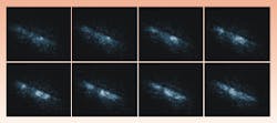
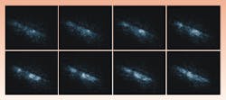
In one experiment, a calcium-ion indicator was inserted into an extracted rat's heart, and the concentration waves of the ions were observed. The calcium ions comprise the information-carrying substance involved in the movement of the heart muscle (see Fig. 1).
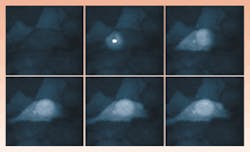
By increasing the laser output, the group has manipulated living cells and has applied external stimulation to the cells using light (see Fig. 2). In the past, such external stimulation has been performed using chemicals, or through strictly mechanical means, such as using glass needles. Researchers found that the concentration of calcium ions increases because of the laser light stimulus, and the transmission of these ions has been directly observed.
The forcing of biological changes via laser stimulation is a first. By applying this technique, it should be possible to control cells remotely using light.
Courtesy O plus E magazine, Tokyo


