MULTIPHOTON IMAGING: Tiny laser makes THG microscopy easy
In third-harmonic-generation (THG) microscopy, third-harmonic light produced by a tightly focused scanned laser beam delineates boundaries inside transparent specimens. The techniquewhich requires the use of ultrafast pulsesmakes possible the construction of three-dimensional images of biological cells without use of an introduced fluorescent dye. Current THG setups, however, rely on Ti:sapphire lasers combined with optical parametric oscillators or amplifiers as a source of femtosecond-scale pulses. Such systems are expensive, complex, and finicky; a THG microscope user versed primarily in biology had better have a friend nearby who is a laser expert.
Researchers at the University of California, San Diego (UCSD; La Jolla, CA) and BioCentrum Amsterdam (Amsterdam, The Netherlands) have made life easier for both biologist and friend by replacing the complex light source with a femtosecond fiber laser that requires no adjustment at all. "[The fiber laser] is environmentally robust," says Jeffrey Squier, director of research. "We found it could handle extensive temperature swings."
Emitting at a spectral peak of 1562 nm, the laser is smaller than a shoebox. It produces a 50-MHz train of 1-nJ, 100-fs pulses at both the fundamental spectral band and visible bands; the visible light must be blocked with an absorptive filter. When focused into a specimen by a microscope objective with a 0.65 numerical aperture (NA), THG is produced with an axial resolution of 7.5 µm; a 1.25-NA objective produces axial resolution of 0.8 µm. The beam is typically raster-scanned across a 250 x 250-µm region, producing a heating rate in the specimen on the order of 3.5 K/s, adequately low for mostbut not allspecimens. A 0.45-NA objective collects the THG light and images it onto a charge-coupled device.
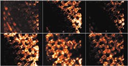
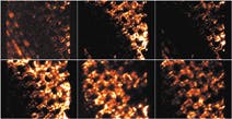
Sections of a THG image of chloroplasts in leaves show ringlike structures (top image). In a nonbiological application, the microscope brightly images an array of 200-nm-diameter spots written within glass with an 800-nm laser and resulting from a change of refractive index (bottom); arrays spaced apart axially can be individually imaged. The THG technique can be used to characterize glass homogeneity, according to Squier.
The UCSD researchers are using their microscope to more-fully understand the contrast mechanisms involved in THG imaging. A future version of the microscope based on a 1.06-µm-emitting fiber laser will transmit better through water, lowering the unwanted heating of biological specimens.
About the Author
John Wallace
Senior Technical Editor (1998-2022)
John Wallace was with Laser Focus World for nearly 25 years, retiring in late June 2022. He obtained a bachelor's degree in mechanical engineering and physics at Rutgers University and a master's in optical engineering at the University of Rochester. Before becoming an editor, John worked as an engineer at RCA, Exxon, Eastman Kodak, and GCA Corporation.


