Optoelectronic Applications: Nondestructive Testing - Laser-based instrumentation sheds new light on old art

Imagine having the opportunity to stand before the Mona Lisa—sans her plexiglass cage, up close and personal, in the basement of the Louvre—and bathe her in coherent blue light, hoping to discover the secrets behind that infamous smile. What minerals and dyes might the pigment contain? What binding agents were used? How much does the final painting differ from the original sketch underneath, and how many layers did it take to create this masterpiece? Did someone—perhaps even Da Vinci himself—at some point alter the original, for reasons we might never know?
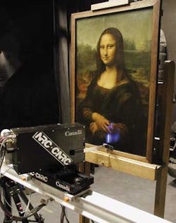
In 2004, researchers from the National Research Council (NRC; Ottawa, ON, Canada) took their 3-D laser scanning equipment to the Louvre in Paris, France, to generate the most detailed analysis to date of the famous painting, providing new information about the wood panel on which it was painted, Da Vinci’s technique, and his original concept for the composition (see Fig. 1). It took more than a year to analyze all the data gathered in two days of scanning, but the results were amazing, according to Francois Blaise, group leader, visual information technology at the NRC Institute for Information Technology. Blaise was instrumental in developing the equipment used on the Mona Lisa and in other 3-D laser-scanning projects at the Louvre, the British Museum (London), and historic sites in Canada and Europe, such as the Acropolis in Athens.
Today, 3-D laser scanning is just one of a number of nondestructive optical techniques that have become indispensable to art historians in museums worldwide. Motivated by a desire to enhance existing conservation techniques and to improve the analysis and authentication of paintings, pottery, statues, tapestries, and related artifacts, art conservators are using fluorescence, infrared, ultraviolet, and Raman spectroscopy; Fourier-transform infrared Raman (FTIR) spectroscopy; coherent anti-Stokes Raman scattering (CARS) microscopy; laser-induced breakdown spectroscopy (LIBS); optical coherence tomography (OCT); and even terahertz imaging to analyze and understand historic art in new and exciting ways.
In 2006, for example, researchers from Harvard University Art Museums (Cambridge, MA) used Raman spectroscopy, scanning electron microscopy, laser desorption-ionization time-of-flight mass spectroscopy, and FTIR to help determine whether three paintings found in a Long Island storage facility in 2002 were actually the work of Jackson Pollock.1 Their analysis of pigments and binding media in the paintings found that many of the materials used were not commercially available until after Pollock died in 1956, casting doubt on the authenticity of the paintings and prompting further study of their authenticity that continues to this day.
“There is a strong trend toward nondestructive testing in art conservation,” says Jens Stenger, associate conservation scientist in the Analytical Lab at the Straus Center for Conservation (part of the Harvard University Art Museums) and a member of the team that analyzed the controversial Pollock paintings. “Several techniques are standard in a museum lab, such as FTIR, x-ray fluorescence spectroscopy, and gas chromatography, but a lot of new technologies are coming into this now and the instruments are getting more affordable, like Raman. Five years ago there were only one or two Raman spectrometers in American museums, but today a lot of museums are buying and using Raman regularly.”
As in forensics, analyzing a piece of artwork typically begins with the most fundamental visualization tool of all: the human eye. A microscopically small sample of pigment is taken from a painting and placed under a standard microscope for examination. A scanning electron microscope is then typically used to look at the same sample with higher magnification. X-ray fluorescence and radiography are also routinely used to gather elemental data. Various spectroscopic techniques are then employed, depending on the desired application, such as Raman spectroscopy for the identification of pigments.
“Raman is really great for pigment identification,” Stenger says. “Recently more and more of the modern synthetic colorants are important because more modern art needs conservation treatment and better understanding from the materials aspect, and Raman is very powerful for identifying modern synthetic dyes (those developed after 1900).” Stenger says his group is currently working on a large project on artist Mark Rothko (1903-1970). “We are interested in how the materials he used changed over his career and how did this coincide with changes in his style,” he says. “Rothko is well known for mixing traditional materials such as rabbit-skin glue with modern synthetic dyes, which is very interesting in terms of art history. Why did he do this, and what effects was he trying to achieve?”
Digging deeper
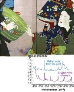
Art historians are now beginning to utilize more sophisticated optical techniques to answer these and other questions about art and those who create it. At the Metropolitan Museum of Art (New York, NY), Marco Leona’s group in the Department of Scientific Research is working with surface-enhance Raman spectroscopy (SERS) to detect natural dyes in archeological and historical textiles, paintings, and other works of art (see Fig. 2).2, 3 Natural organic products historically used as dyes or pigments are often fluorescent under normal Raman measurement conditions. In addition, the amount of dye present on works of art is minimal, requiring extremely sensitive analytical techniques. The enhancement of the Raman signal and the quenching of the background fluorescence resulting from the adsorption of dye molecules on metal nanoparticles in SERS solve the problems encountered when studying dyes by traditional Raman spectroscopy.“Raman has entered our field in the last five years because it is more inexpensive and available and it can be used with a microscope,” Leona says. “But if you are interested in studying natural organic colorants, it is very difficult to do with Raman spectroscopy because they are very fluorescent and always used in very small amounts with many interfering materials. SERS became interesting because it is highly sensitive down to single molecules in the right conditions, and you can suppress the fluorescence and enhance the Raman response. You still have to remove a sample, but it can be 100 times smaller than if doing high-performance liquid chromatography.”
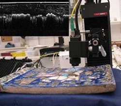
Optical coherence tomography is also gaining favor in art conservation circles. Scientists at the British Museum have been investigating the use of OCT for in situ surface and subsurface analysis since 2004; they have worked with time-domain and Fourier-domain OCT (930 and 1330 nm) to determine, for example, whether a painting has been painted over for some reason and they have conducted systematic studies of the spectral transparency of historic artists’ paint, identifying the spectral region best suited to OCT imaging of paint layers.4 David Saunders, keeper of the Department of Conservation, Documentation and Science, at the museum, says that noninvasive point-based techniques such as OCT provide a nice complement to IR and UV spectroscopy and x-ray fluorescence. These techniques provide insight into the gross structure and location of different materials across the surface and subsurface; OCT can then be used to noninvasively obtain stratigraphic information across the object.
“We are looking at what you can determine using OCT to investigate the surface and subsurface of objects, and what is promising is the ability to see if the layer structure at the sample site holds across the whole area,” Saunders says. “We are also looking at which wavelengths and methods for processing the signal give different types of information about the object, and whether we can move beyond it as simply an imaging tool to get more physical properties information about the subsurface layers and the nature of the material.”Stenger and colleagues at Harvard have collaborated with researchers from Massachusetts Institute of Technology to evaluate swept-source Fourier-domain OCT for nondestructive analysis of gold punchwork and underdrawings in Renaissance panel paintings.5 Their research concluded that this technique is attractive for art conservation because of its unique combination of ultrahigh imaging speeds, large ranging depths, and 1300 nm wavelength, which enables enhanced visualization of deeply buried structures due to reduced scattering.
“We used OCT to get detailed shots of the underdrawings because carbon (from the original pencil or charcoal) absorbs very well in the IR,” Stenger says. “With OCT we can obtain very detailed images to characterize the carbon-based material used, and also obtain depth information that determines in which layer of the painting the underdrawing is.”
Optical coherence tomography may also play a role in improving the restoration of paintings. Researchers from Nicolaus Copernicus University (Torun, Poland) have been using spectral-domain OCT to monitor Er:YAG laser ablation of varnish layers.6 Their goal is to determine the optimal laser fluences for cleaning and removing old varnish without damaging the underlying artwork in the process.
“There is a lot of controversy about laser cleaning of paintings because the varnish is bound with much the same organic material as the paint underneath,” Saunders says. “OCT potentially offers some advantages here, although there is still a lot of basic research that needs to take place before people will be comfortable with laser cleaning for paintings.”
Terahertz imaging is another technique being investigated for art restoration. Researchers from the University of Michigan and the Louvre recently utilized terahertz technology from Picometrix to reveal murals hidden beneath coats of plaster and paint in centuries-old buildings in France.7 The technology is expected to find further application in Europe, where many famous artworks—such as DaVinci’s “The Battle of Anghiari”—are believed to have been painted or plastered over to protect them from falling into enemy hands. In March the researchers took the equipment to Vif, France, to help archeologists examine a mural discovered recently behind five layers of plaster in a 12th century church.
“Initially there were crumbling plaster walls that revealed small section of murals and they were trying to remove some of the plaster without damaging the art underneath,” says John Whitaker, researcher scientist and adjunct profession of electrical engineering and computer science at the University of Michigan. “Now we are using the terahertz first to help them see what is there and to image parts of the wall for them. It is not a big church, but we think it is predominantly covered with these murals. If we can image this before they remove the plaster, then they will know that if there is damage during the removal process, they can restore the original murals based upon our images.”
REFERENCES
1. R. Kennedy, “Harvard Analysis Casts Doubt on Works Said to Be Pollocks,” New York Times, Jan. 30, 2007.
2. M. Leona, J. Raman Spectroscopy 37, 981 (2006).
3. K. Chen, M. Leona, T. Vo-Dinh, Sensor Review 27/2, 109 (2007).
4. H. Liang et al., Proc. of SPIE 6618, 661805 (2007).
5. D. Adler et al., Optics Express 15/24, 15972 (2007).
6. M. Gora, P. Targowski, A. Rycyk, J. Marczak, Laser Chemistry, doi:10.1155/2006/10647.
7. J.B. Jackson et al., Optics Communications 281, 527-532 (2008).
About the Author
Kathy Kincade
Contributing Editor
Kathy Kincade is the founding editor of BioOptics World and a veteran reporter on optical technologies for biomedicine. She also served as the editor-in-chief of DrBicuspid.com, a web portal for dental professionals.

