Advanced components raise Raman sensitivity
Advanced components raise Raman sensitivity
Raman instruments with near-infrared diode lasers and enhanced CCD cameras can minimize problems of sample fluorescence and low sensitivity.
Harry Owen and Will K. Kowalchyk
Compact, integrated charge-coupled-device (CCD)-based spectrometers are making possible a renaissance in Raman spectroscopy. Rapid growth is happening in all major application areas--basic research, industrial R&D, and on-line process control. Advances in both laser and CCD technologies are bringing about a second generation of instruments with improved performance at longer excitation wavelengths that eliminate problems of sample fluorescence. Preliminary data from a pilot US Environmental Protection Agency (EPA) groundwater pollution study demonstrate the utility of sensitive, fluorescence-free Raman spectroscopy.
Compared to other spectroscopic techniques, Raman offers several critical advantages. For example, it can be performed with ultraviolet (UV), visible, or near-infrared (NIR) excitation. This flexibility allows use of traditional optical systems ranging from fiberoptic probes to confocal microscopes for sampling. Most Raman spectroscopy requires no special sample preparation, allowing in situ measurements on samples varying from living cells to refinery streams. Also, unlike NIR spectroscopy, Raman spectra consist of shar¥peaks (fundamental vibrations), so a sample concentration often can simply be derived from a peak area.
Raman scattering, however, is a low-probability event resulting in low signal levels. For this reason, it formerly required complex and expensive instrumentation and was generally unsuited to anything except pure research. This situation changed with the advent of CCD-based Raman spectrometers, which deliver the turnkey convenience associated with other types of spectrometers. The use of compact, high-power lasers with holographic notch filters and transmission gratings in these instruments enables the efficient generation and detection of the inherently weak Raman signal with excellent signal-to-noise (S/N) characteristics.
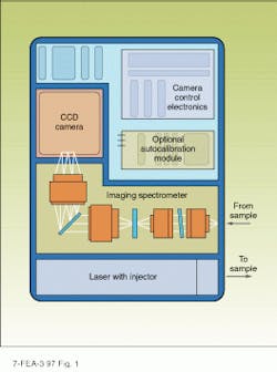
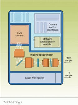
Examples of CCD-based Raman spectrometers include the Kaiser HoloLab and HoloProbe spectrometers (see Fig. 1). The excitation source is either a frequency-doubled diode-pumped Nd:YAG laser emitting at 532 nm or a stabilized diode laser with output at 785 nm. Because of its short wavelength and higher output power, the Nd:YAG laser is preferred when maximum sensitivity is required and no sample fluorescence is present.
Laser light is coupled into a fiber for delivery to the sample, which can be hundreds of meters from the spectrometer if desired. Holographic optics in the probe head eliminate any Raman signal from the fiber itself. The Raman scattering is collected and returned via a second fiber. A holographic notch filter removes the Rayleigh scattered laser light before the remaining Raman signal is dispersed in an f/1.8 axial transmissive spectrograph.
The dispersed light is detected by a thermoelectrically (TE) cooled CCD camera, with typical configuration of 1024 ¥ 256 pixels. In addition to low-noise detection of low photon fluxes, a cooled CCD enhances the S/N ratio and reduces data-acquisition times. A further throughput advantage is achieved with the HoloPlex grating that enables simultaneous acquisition of the entire spectrum (0-3000 cm-1 ) with no scanning (see Laser Focus World, Oct. 95, p. 95). With several collection fibers stacked vertically at the input of the spectrograph, a single CCD camera can detect several dispersed spectra simultaneously. In a chemical plant or refinery, this allows a one instrument to monitor several points and control an entire process.
Long-wavelength excitation
Many analytical applications require quantitative detection of low concentrations of chemicals that may be relatively weak Raman scatterers. These environmental and process control applications often include other species in the sample that fluoresce when excited with visible light. Because fluorescence can be u¥to 108 times stronger than Raman scattering, even trace levels of a fluorescent species can contribute a significant amount of background light. This lowers system S/N and can preclude accurate quantitative analysis of the Raman spectrum. In the worst case, the Raman signal may be completely swamped by the background fluorescence. Many samples with biological or organic content will fluoresce with visible excitation, which has been a major factor limiting the use of visible-wavelength Raman spectroscopy for pharmaceutical and environmental studies and in the food industry.
Sample fluorescence can be minimized, however, by using a near-IR laser source, because long wavelengths cannot excite most molecules to their upper electronic states (where fluorescence originates). But which near-IR laser? The fundamental output of a Nd:YAG laser at 1064 nm is not suitable for CCD-based Raman instruments because the Stokes-shifted Raman signals will fall outside the wavelength detection range of the silicon CCD pixels. A titanium:sapphire laser could provide the ideal 750-800-nm wavelength, but it is currently too complex, bulky, and expensive to integrate into a turnkey Raman spectrometer. A diode laser is a suitable compact near-IR laser source.
Stable, high-power laser diode
Diode lasers controlled with an external grating can achieve narrow line output, with the wavelength stability and transverse mode characteristics necessary for quantitative Raman measurements. Until recently, external-cavity diode lasers were limited to maximum output powers in the 20-25-mW range. This power level is too low for quantitative applications because Raman signal intensities are proportional to the fourth power of the excitation frequency (photon energy).
So although long-wavelength excitation may eliminate fluorescence, it also significantly lowers the Raman signal level. Our goal was to build an instrument with a net increase in sensitivity. We turned to laser manufacturer SDL (San Jose, CA) for an OEM version of its model 8530 external-cavity diode laser emitting at 785 nm. According to Milan Zeman, product manager for scientific products at SDL, the company spent the past few years developing a Raman source. Key to the laser performance is a tapered amplifier diode. The SDL-designed chi¥has a tapered gain region that emits hundreds of milliwatts in a diffraction-limited beam. In the HoloProbe and HoloLab series, we limit the output of the 8530 laser to 250 mW and add a holographic bandpass filter module to eliminate the super-radiance sometimes associated with high-power diode-laser output.
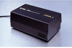
In addition to output power, laser wavelength stability is important, because peaks due to Raman scattering do not occur at fixed wavelengths, but rather at fixed frequency shifts relative to the laser source. A second-generation external cavity stabilizes laser output in the model 8530 diode laser (see Fig. 2). [See Laser Focus World, May 1995, p. 117 for discussion of an early prototype of this laser--Ed.] Laser wavelength is set and fixed at 785 nm according to our specification, which allows easy interchangeability in the process-control environment. With the entire laser cavity temperature-stabilized, the wavelength of this sealed unit is stable to 0.1 nm, with a linewidth of 10 GH¥(0.02 nm). An integrated 30-dB optical isolator in the output beam provides immunity to feedback reflections, and the output is a diffraction-limited 1.5-mm-diameter circular beam that is efficiently coupled into fiber.
High-sensitivity CCD camera
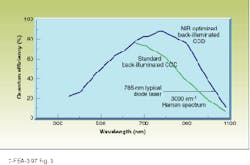
Many useful spectral features occur at large Raman shifts (2500-3100 cm-1), most notably those associated with C-H and S-H stretching vibrations. Consequently, the spectrometer must be able to efficiently detect light that is Stokes-shifted 3000 cm-1 from 785 nm (which corresponds to 1026 nm), so we needed a CCD camera with reasonable sensitivity out to at least 1030 nm. The quantum efficiency (QE) of silicon-based photo detectors decreases rapidly above 900 nm--conventional CCDs have a QE of only 7% at 1000 nm and 3% at 1050 nm; back-illuminated devices have somewhat higher QE values: 19% at 1000 nm and 6% at 1050 nm.
Princeton Instruments (Trenton, NJ) designed a custom TE-cooled camera (it has a CCD chi¥optimized for high QE at long wavelengths) for exclusive use within our spectrometer. Roy Howard, spectroscopy specialist at Princeton, explains, "Because silicon is relatively transparent to longer wavelengths, the average absorption depth at 1000 nm is 100 µm. But in a typical CCD, the depletion zone, that is, the region from which photogenerated charge is collected, is only 5 µm." Princeton used a novel back-illuminated CCD design using high-resistivity silicon to increase the depletion region to 20 µm.
A proprietary antireflection surface treatment defeats multiple internal reflections in the silicon. Howard notes, "This is important, because silicon has a very high refractive index (3.5) at 1000 nm. Without this treatment, conventional back-illuminated CCDs produce an etalon effect--fringes are superimposed on the response curve of the CCD. With this treatment, no such fringes are seen." In the back-illuminated device, the QE is more than 39% at 1000 nm and is still 23% at 1050 nm (see Fig. 3).
Groundwater monitoring
How does this technology relate to real applications? The EPA National Exposure Research Laboratory (Athens, GA) bought one of the first of these instruments to evaluate NIR Raman spectroscopy for the in situ measurement of low-level organic contaminants in groundwater. An important potential application involves measuring MTBE (methyl tertiary-butylether) in contaminated groundwaters. Contamination of groundwater with MTBE is a highly publicized issue in Santa Monica, CA, and elsewhere. The chemical is used in many states as an additive to produce oxygenated gasoline, which results in less pollution when combusted. Typical MTBE concentrations in gasoline are between 11% and 15%.
Large quantities of gasoline are stored in underground tanks, from which slight leakage is inevi ta ble. Measurable amounts of MTBE have begun to turn u¥in groundwater samples, partly be cause MTBE is more mobile than other gasoline constituents and signi ficantly more miscible with water. Tim Collette, re search chemist at the EPA, says, "Our ultimate goal would be to sample many points in the field with real-time in situ measurements, using a fiber probe and cone penetrometer. This would allow us to ma¥the `plume` of MTBE as it spreads in the water table. It might also allow us to monitor the effectiveness of remediation."
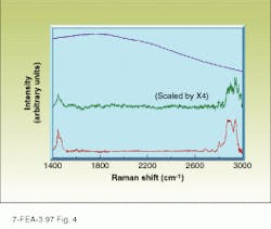
Some of the strongest MTBE Raman peaks are C-H stretches at 2700-3000 cm-1, and groundwater has trace amounts of humic acid, fulvic acid, and other bio-organics that could cause fluorescence problems with short-wavelength excitation. We obtained several spectra of MTBE (2 ¥ 103 ppm) in water with 10 ppm fulvic acid (see Fig. 4). The first was obtained with 532-nm excitation and shows a level of background fluorescence high enough to completely obscure the Raman spectrum. The second illustrates the effects of switching to a high-power 785-nm laser--fluorescence is eliminated but the Raman S/N ratio is not particularly good.
The third spectrum was run using the new instrument with both 785-nm excitation and the near-IR optimized CCD, and it clearly shows the boost in S/N ratio. The first two spectra were recorded in Kaiser`s applications laboratory, the third, at the EPA laboratory in Athens, GA. Data-acquisition time was 60 s with 10 accumulations in each case. Collette estimates current detection limits with the new instrument at about 10-3 molar. Although he would prefer 10-4 molar or better, he believes that a novel sample enrichment probe he is developing might be able to achieve better limits.
Raman spectroscopy is now successfully competing in the $3 billion analytical instrumentation market with separation methods such as gas chromatog raphy and high-pressure liquid chromatography. While Raman is not yet able to reach the high sensitivity levels of these instruments, it is often less time-consuming to perform and offers more flexible implementation, with potentially significant cost-of-ownershi¥advantages. Advances in Raman sensitivity could lead to the technique becoming the preferred choice in an increasing number of analytical and process-monitoring applications. o
Disclaimer
Mention of trade names or commercial products does not constitute endorsement or recommendation for use by the US Environmental Protection Agency.
FIGURE 1. Diode laser or frequency-doubled Nd:YAG laser provides excitation in CCD-based Raman spectrometer such as the HoloProbe.
FIGURE 2. The SDL 8530 laser produces several hundred milliwatts at 785 nm from a compact package, making it ideal for integration into a Raman spectrometer.
FIGURE 3. Combination of dee¥depletion, back-illumination, and antireflection coatings dramatically boosts CCD response at long wavelengths.
FIGURE 4. Three different instruments were used to record MTBE Raman spectra under identical conditions. The sample is 2 ¥ 103 ppm MTBE in water with 10 ppm fulvic acid. Upper trace was made with 532-nm excitation and front-illuminated CCD detection. Middle trace was made with high-power 785-nm excitation and a front-illuminated CCD. Lower trace resulted from high-power 785-nm excitation and a NIR-optimized CCD. Water background has been subtracted from all spectra.
HARRY OWEN is director of new business development and WILL K. KOWALCHYK is applications scientist, Laser Spectroscopy Products, Kaiser Optical Systems, 371 Parkland Plaza Rd., Ann Arbor, MI 48103.
