Increased pressure to reduce health-care costs coupled with a growing trend toward managed care have created new opportunities for medical diagnostics. In the past few years attention has turned toward light-based technologies, primarily because of their ability to noninvasively image and analyze tissue function and structure in real time or nearly real time. Optical diagnostic techniques offer the potential to not only enhance existing capabilities, but to eliminate the need for physical biopsies.
The development of commercial light-based diagnostic tools has been slow, primarily due to the unpredictable nature of photon absorption and scattering in tissue. In addition, although optical diagnostic techniques are capable of gleaning more information from tissue than conventional techniques, they also require greater analytical capabilities. Fluorescence spectroscopy, for example, can be used to detect certain metabolic changes representing early tumor growth; however, this information must be analyzed before any diagnosis can be rendered. Meanwhile, the demand for real-time imaging techniques continues.
Near-infrared (NIR) lasers operating between 700 and 900 nm are improving our understanding of how light propagates in biological material. Maximum light penetration into tissue occurs in this spectral window, enhancing researchers’ ability to characterize optical properties of tissue and correlate them with physiological processes. These lasers also have been instrumental in pushing the first optical diagnostic tools to the brink of commercialization and in setting the stage for several second-generation devices.
Fluorescence vs. Raman spectroscopy
Current optical-diagnostic research generally falls into two categories: spectroscopy (fluorescence and Raman) and imaging (including photon migration and optical tomography). Clinical applications of both have been pursued for several years; those applications involving fluorescence spectroscopy have progressed furthest in terms of practical realization.
In medicine, spectroscopy is principally used to probe specific molecules and the environment around those molecules or to measure time-dependent variations of photon absorption and scattering in large volumes of tissue. Fluorescence spectroscopy is more suited to gathering metabolic or structural data, while Raman spectroscopy is used for more specific purposes, such as studying protein structures.
“At the molecular level, I don’t think we fully understand why Raman spectroscopy is more specific than fluorescence spectroscopy,” says Rebecca Richards-Kortum, associate professor of biomedical engineering and electrical and computer engineering at the University of Texas (UT, Austin, TX). “With fluorescence, we are limited to a penetration depth of 300 µm, and only 20 or so molecules contribute information. With Raman, we have more of a fingerprint perspective; we can look as deep as 1 mm when working in the near-infrared.”
A number of researchers are working to better understand the distinctions between fluorescence and Raman spectroscopy in diagnostic samples and to apply them accordingly. For example, Richard McCreery’s group at Ohio State University (Columbus, OH) is studying what contributes to the Raman spectrum in breast cancer; Arjun Yohd, associate professor of physics at the University of Pennsylvania (Philadelphia, PA), and colleagues are combining fluorescence spectroscopy and NIR imaging to detect metabolic changes in the brain; and the Richards-Kortum group is using both fluorescence and Raman spectroscopy to detect cervical precancer.
“We have a collaborative project with the M.D. Anderson Cancer Center (Houston, TX) to develop a real-time diagnostic tool for cervical precancer,” Richards-Kortum explains. “We are trying to improve colposcopies (in which a dissecting microscope is used to detect cancerous cells on the cervix after an abnormal Pap smear), which are not very specific and require a biopsy and at least one follow-up visit.”
Using funding from LifeSpex (Houston, TX (see “First optical-mammography system in clinical trials,” below), the Richards-Kortum group used a nitrogen-pumped dye-laser (at 337, 380, and 460 nm) for fluorescence spectroscopy. The group has studied more than 250 patients with the instrument. Results have revealed the ability to differentiate tissue with 20% greater specificity than currently possible with conventional methods. The UT group recently received funding from the Whittaker Foundation to expand to multicenter trials and is now also investigating Raman spectroscopy. In addition, LifeSpex has a Cooperative Research and Development Agreement with Sandia National Laboratories (Albuquerque, NM) to build a more rugged fluorescence system.
Faster analysis
Fluorescence and Raman spectroscopy are also being used to distinguish cancerous from noncancerous tissue in the mouth, esophagus, and colon. Researchers at the City College of New York Institute for Ultrafast Spectroscopy and Lasers (New York, NY) have constructed prototypes of three lamp-based products—the CD scanner, CD ratiometer, and CD map—that are based on fluorescence techniques developed and patented by institute director Robert Alfano, M.D.
“We excite with lamps, not lasers, because of the tunability,” Alfano says. For example, the CD scanner uses a fiberoptic delivery/collection system to measure fluorescence as a function of wavelength. In mouth and esophagus studies at Sloan-Kettering Institute (New York, NY), the same instrument was used to excite tryptophan, collagen, elastin, and NADH—a key molecule for monitoring metabolic changes. “Everything is measured relative to the different peaks found in tryptophan, so we don’t have to worry about intensities,” Alfano says.
The CD ratiometer uses a filter to quickly read ratios at various wavelengths, while the CD map uses a video system to study ratios at various wavelengths and points in a field. Studies are continuing at Sloan-Kettering and Columbia-Presbyterian Medical Center (New York, NY), and investigational-device-exemption applications are pending. Much of the development work was funded by Mediscience (Cherry Hill, NJ).
Alfano has also patented a diode-laser-based Raman fluorescence apparatus for histology. “We have shown that a Raman device can isolate cancers in the cervix, but [the signal] is very weak, so it is more difficult to analyze an image,” he says. “Raman is the next generation, but there is still a lot of work to be done to quantify it as a useful tool.” The group is now working to develop faster and more reliable algorithms for data analysis, hoping to bring diagnostics to the 15-s regime.
Under the direction of Michael Feld, researchers at the Massachusetts Institute of Technology Laser Biomedical Research Center (Cambridge, MA) are enhancing data analysis with a video system and signal spectrometer. The group has also developed a portable, diode-laser-based NIR Raman spectrometer to analyze blood chemistry. The system is currently in clinical trials and has been used in patients to detect early breast and bladder cancers, sarcomas, and atherosclerosis in coronary and peripheral arteries.
“We adapted a device based on nonimaging optics that allows you to extract 12 times more light than is possible with an optical fiber,” Feld says. “We are trying to measure things at an early stage to eliminate breast biopsies and other invasive diagnostic techniques.”
Photon migration
Early cancer detection is also the focus of several optical-imaging studies. Optical imaging is generally used to gather structural information about tissue and to image tumors or other localized tissue masses; more specifically, various frequency- and time-domain techniques can measure tissue hemoglobin, blood volume, water content, and cellular structure in superficial tissue layers (less than 1 mm deep), while techniques such as optical tomography probe several centimeters into tissue to produce images from certain emitted photons.
Here again, NIR lasers are contributing to the development of practical devices. According to Enrico Gratton of the Laboratory for Fluorescence Dynamics at the University of Illinois (Urbana-Champaign, IL), although NIR optical imaging offers real-time data access, portability, and cost-effectiveness, image reconstruction is complicated by the diffusive character of light propagation in optically heterogeneous scattering tissue.
This issue has spawned a growing number of NIR photon-migration studies. For example, Bruce Tromberg, M.D., associate professor of physiology and biophysics at University of California, Irvine, and a faculty member at Beckman Laser Institute (Irvine, CA), is studying the frequency dependence of photon-density waves in tissue to determine optical properties such as oxygen physiology, blood volume, and water content. These data can then be interpreted quantitatively at various wavelengths and can be correlated to certain physiologies, such as the ratio of oxygenated to deoxygenated blood. “With this information, we can then further probe tissue physiologies, determine dosimetries of laser therapies, find malignant transformations or tumors, monitor muscle activity, and detect traumatic injuries to tissue,” he says. “It is not imaging, but localized spectroscopy.”
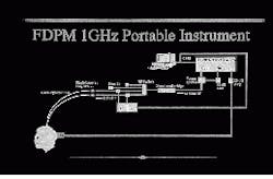
Tromberg and his colleagues have developed a hand-held, 1-GHz frequency-domain photon-migration (FDPM) instrument for measuring optical properties of tissue such as oxygen physiology, blood volume, and water content (see Fig. 1). Though not originally intended for commercialization, the device has attracted commercial interest; at this point, the Beckman Institute has protocols to study uterine and breast tissue and to determine photodynamic-therapy dosimetries.
Tromberg is also collaborating with Arjun Yohd and Britton Chance, professor emeritus of biochemistry, biophysics, and radiology at the University of Pennsylvania (Philadelphia, PA), to combine their imaging expertise with the data-collection capabilities of FDPM technology. “Our approach is to develop hand-held sensors for making these measurements one spot at a time,” Tromberg says. “Yohd is interested in forming images and has developed a very good method for this. What we have is a unique way of finding data that could be used to form those images.”
Diffusing light
According to Yohd, whose group works closely with Britton Chance, most of the optical-imaging research at the University of Pennsylvania involves NIR diffusing photons. “Over time, we have gone from looking at simple properties of the waves to developing functional imaging devices,” he says.
Some of this work has found its way into a fiber-coupled, diode-laser-based “regional brain imager” being commercialized by a young Philadelphia company. Yohd says brain studies are promising because light in the near-IR penetrates the skull much more effectively than, say, ultrasound. In addition, he says, measurements of oxygen dynamics are facilitated because optical spectra are instrinsically sensitive to blood oxygenation, whereas magnetic resonance imaging detects blood indirectly.
“Some modulated light continues to oscillate even after it has scattered, and this is what is detected,” Yohd says. “Thus, you can reconstruct an image of the volume of tissue you are looking at and determine such things as blood oxygenation and metabolism.”
Applications of this technology can be expanded, he adds, by considering temporal fluctuations of emitted light to see more cell motion or adding contrast agents to compare lifetime changes. “Suppose the metabolism is high in one region compared to the others,” he says. “This could be a sign that something is rapidly growing there, such as a tumor. Or we could find that a certain medication disturbs delivery of oxygen to a baby’s brain. We then just need to determine why.”
At the Laser Biomedical Research Center, Feld’s group has achieved good results with photon migration and optical imaging. “We are interested in imaging disease throughout the body and diagnosing precancer in mucosal surfaces using fluorescence-based photon migration,” he says. For example, using a specially designed endoscope, the group has obtained some “very promising” diagnostic results for diseases of the colon. “But it takes a lot to make a practical device,” he adds.
At the Institute for Ultrafast Spectroscopy, Alfano’s staff is studying optical breast-imaging techniques using intralipids (a highly scattering, milk-like substance meant to simulate dense tissue), a chromium-doped forsterite laser (tunable from 1.1 to 1.3 µm), time and space gating, and Fourier filtering.
“Because near-IR light gets through the body, we are working on model systems to see how light propagates through highly scattering media,” Alfano says (see Fig. 2). “We want to see the image and find the key wavelength that distinguishes cancer from noncancerous tissue.” That wavelength should be somewhere between 800 and 1200 nm, he adds. “I believe water absorption is going to be a key factor in developing a successful breast imager.” In addition, he says, 10 ps appears to be fast enough for gating out “snake light” (those photons that propagate through tissue first) and achieving a clear image.Other techniques
One of the most promising areas of development in optical diagnostics is fluorescence-lifetime imaging (see Laser Focus World, May 1992, p. 60). According to Joe Lakowicz, director of the Center for Fluorescence Spectroscopy at the University of Maryland (Baltimore, MD), one important reason for this is that fluorescence-lifetime techniques are insensitive to real-world factors such as sample turbidity, optical-surface contamination, or variations in optical alignment. In addition, intensity-based measurements often cannot account for the erratic nature of light as it travels through dense media.
Fluorescence-lifetime techniques, however, have been shown to circumvent the unpredictable absorption and scattering properties of tissues, says Eva Sevick-Muraca, assistant professor of chemical engineering at Purdue University (West Lafayette, IN). Using an argon-ion-pumped Ti:sapphire-laser system, she and her colleagues at Purdue’s Photon Migration Laboratory have shown that when excitation and fluorescent photons migrate similarly, the relationship between the phase-shift and amplitude-modulation values and the re-emitted excitation light provides direct tissue information independent of tissue optical properties. “Our approach could be used to conduct endoscopic measurements of endogenous or exogenous fluorescence lifetime without the scattering and absorption changes that may occur with pathophysiology,” she says.
More recently, Purdue researchers have demonstrated that fluorescence-lifetime spectroscopic imaging can be used to diagnose disease in deep tissue by detecting specific biochemical changes. According to Sevick, images of fluorescent lifetime can be reconstructed from frequency-domain measurements of excitation and fluorescent phase shifts; this can, in turn, provide a map of biochemical conditions that may signal disease.
Using some of the same principles, Enrico Gratton’s group has developed a scanning lifetime-fluorescence microscope that relies on stimulated emission to produce ultrahigh-resolution images. According to Gratton, this device offers significant advantages. The cross-correlation signal produced by overlapping the pump and probe lasers results in an axial-sectioning effect similar to that in confocal and two-photon excitation microscopy and improved spatial resolution compared to conventional one-photon fluorescence microscopy. The low-frequency cross-correlation signal allows lifetime-resolved imaging without the need for fast photodetectors. Medical-device manufacturer Carl Zeiss is now studying the viability of this approach for imaging breast tumors.
First optical-mammography system in clinical trials
Imaging Diagnostic Systems (IDS, Ft. Lauderdale, FL) is planning to conduct clinical studies of what appears to be the first commercial optical-mammography system. According to the company, the CT Laser Mammography system uses tomographic scanning geometry, a Ti:sapphire laser (this may change) and detectors to produce slice-plane images and cranial-caudal and lateral-medial image projections.
Although several optical-imaging experts question how IDS could be this far along in the development of an optical-imaging system, IDS has been awarded several patents, successfully went public last year, and has exhibited at the 1994 and 1995 meetings of the Radiological Society of North America. In addition, the IDS optical-mammography system was featured on television on the Phil Donahue Show last November.
Other optical diagnostic products that are being developed:
- LifeSpex (Houston, TX), formerly Patient Technologies Inc. (Albuquerque, NM), is working with researchers at M. D. Anderson Cancer Center (Houston, TX) to develop a fiberoptic fluorescence spectroscopy probe and related products for real-time detection of cervical precancer.
- Biophotonics (Cambridge, MA) has developed a laser-based fluorescence-spectroscopy device called the Colpo Probe. Initial testing at Beth Israel Hospital (Boston, MA) produced “promising results”; clinical trials are expected to begin this year.
- Fonet (Clearwater, FL) is in Phase 2B clinical studies of a lamp-based fiberoptic spectral analyzer developed at Los Alamos National Laboratory (Los Alamos, NM) for optical biopsies. Because it can measure spectra only on tissue it touches, the device is expected to be used primarily for superficial cancers of the colon, bladder, cervix, uterus, skin, and eye.
- Mediscience (Cherry Hill, NJ), which is commercializing optical-tomography technology developed at the Institute for Ultrafast Spectroscopy and Lasers (New York, NY), signed a major agreement with General Electric late last year to further develop this technology.
- Humphrey Instruments (San Leandro, CA), a division of Carl Zeiss, is commercializing an optical coherence-tomography system developed by researchers at the Massachusetts Institute of Technology (Cambridge, MA), Lincoln Laboratory (Cambridge, MA), and the New England Eye Center (Boston, MA).
- Novametrix Medical Systems (Wallingford, CT) and Lawrence Livermore National Laboratory (Livermore, CA) have a $1.8 milion CRADA to develop laser-based sensors to detect and monitor oxygen, alcohol, and other compounds in blood.
- A start-up company called NIMS (Philadelphia, PA) is working with researchers from the University of Pennsylvania on a diode-laser-based “regional brain imager” that can detect metabolic changes by noting wave fluctuations in modulated light and measuring the average absorption in tissue. Potential applications include tumor detection.
- ISS (Urbana, IL), a commercial spin-off from the University of Illinois (Urbana-Champaign, IL), reportedly is developing spectral fluorometers and an oxymeter that uses photon-migration techniques to study tissue physiology and scattering.
- A HeCd-laser-based fluoresence diagnostic system developed by Xillix (Vancouver, BC, Canada) for early detection of lung cancers has been in clinical trials in Canada since 1991 (see Laser Focus World, Feb. 1994, p. 49).
- Star Medical (Pleasanton, CA) has developed a diode-laser-based system to determine the depth of a burn by using indocyanine green dye and fluorescence to measure blood flow. The device is in clinical studies at Shriners Burn Institute (Boston, MA) and is currently serving as the diagnostic component of a burn-debridement system being developed at Wellman Laboratory of Photomedicine (Boston, MA).

