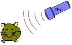
Terahertz radiation affects gene expression in mouse stem cells
Los Alamos, NM--While the use of terahertz radiation is becoming more and more common in medical, military, security, and research applications, few studies have been done on the effects of terahertz radiation on living things. Now, a team of researchers led by Los Alamos National Laboratory has examined the cellular response of mesenchymal mouse stem cells exposed to terahertz radiation, finding some clear effects.1
Two terahertz sources were used in the experiment: a pulsed broadband source centered at 10 THz, and a continuous-wave 2.52 THz molecular-gas laser. The broadband radiation was created by directing ultrafast fundamental and second-harmonic laser light into a pressurized atomic gas, creating terahertz pulses with energies of 1 μ and durations of about 35 fs, with a repetition rate of 1 kHz.
The group determined that temperature increases from the radiation were minimal, and that heat-shock protein expression was unaffected. However, the expression of certain other genes showed clear effects from terahertz irradiation. As the researchers describe in the September issue of the Optical Society’s (OSA) open-access journal Biomedical Optics Express, the stem cells exposed to prolonged broadband terahertz radiation showed specific changes in cellular function closely related to the expression of Adiponectin, GLUT4, and PPARG genes.
The researchers believe further investigations involving a large number of genes and variation in terahertz-radiation characteristics and exposure duration are needed to generalize their findings. They also say that more direct experimental investigations of terahertz radiation’s ability to induce specific openings of the DNA double strand are needed to fully determine how terahertz radiation may work through DNA dynamics to influence cellular function.
The team worked in collaboration with the Center for Integrated Nanotechnologies, a U.S. Department of Energy, Office of Basic Energy Sciences user facility at Los Alamos and Sandia National Laboratories, and with Harvard Medical School, and Beth Israel Deaconess Medical Center.
REFERENCE:
1. Boian S. Alexandrov et al., Biomedical Optics Express, Vol. 2, No. 9, 1 Sept. 2011.
About the Author

John Wallace
Senior Technical Editor (1998-2022)
John Wallace was with Laser Focus World for nearly 25 years, retiring in late June 2022. He obtained a bachelor's degree in mechanical engineering and physics at Rutgers University and a master's in optical engineering at the University of Rochester. Before becoming an editor, John worked as an engineer at RCA, Exxon, Eastman Kodak, and GCA Corporation.