Laser light measures ocular microtremors
If the eye is the window to the soul, it is the eye's motions that make it so: observable movements of the eye help to reveal emotions, moods, and even thoughts. Less familiar to most people is the fact that other, much smaller, eye movements can provide a window to the functioning of the brain itself. Researchers at St. James's Hospital (Dublin, Ireland) have developed an optical technique to measure these microscopic motions to a high degree of accuracy. The noncontact technique, which is based on a speckle interferometer that detects the movement of the sclera (or white) of the eye, can potentially lead to small, headset-sized measurement devices.
Of all the human senses, vision is the most closely connected with the brain—in fact, the developing eye of the human embryo forms as an outpocketing of the brain. Layered upon the large eye motions that are a result of conscious behavior or dreaming are three types of microscopic motions: drift, microsaccades, and ocular microtremor (OMT). All three are influenced by brainstem function. Drift is a continuous irregular wandering of the eye with a range of about 1 arc min, while microsaccades are small darting motions of about 5 arc min in amplitude that occur a couple of times a second. Ocular microtremor is more complex, combining interludes of apparent randomness with bursts of periodic motion. Spectral analysis of normal eye movement shows frequencies due to OMT that span 40 to 100 Hz and peak at about 80 Hz. Amplitudes of OMT fall between 4 and 30 arc sec (lateral displacements of 0.2 to 2 (m).
A link in the brainstem
Ocular microtremor may be of great significance to medical researchers. Analysis of the form and frequency of OMT may help identify various clinical conditions, such as Parkinson's disease, as well as monitor depth of anesthesia. Even more important, the existence or absence of OMT may give an immediate answer as to whether a person in a coma has undergone brainstem death. When OMT does exist during coma, an increase in its frequency indicates the patient is recovering. According to Gerard Boyle, one of the researchers at St. James's Hospital, this link may occur because centers in the brainstem that control eye movement lie close to the so-called reticular formation, which is associated with arousal states such as coma, sleep, and brainstem death.
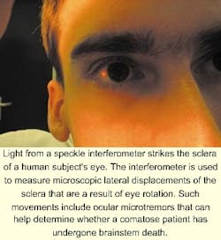
Existing investigations of OMT have relied on the use of eye-contacting piezoelectric probes that require anesthesia of the eye and can induce eyelid spasms that make measurement impossible. To avoid these problems, Boyle and his colleagues built a phase-modulated speckle interferometer that measures the lateral displacement of a spot on the sclera 35° away from the eye's optical axis. In the prototype instrument, two beams of 633-nm light irradiate the sclera from opposite 30° angles, with one of the beams modulated at 15 kHz (see figure). A nonimaging photodetector captures the signal, which is sampled at 16 kHz and digitally demodulated. Laser light levels are kept well below safety limits.
The researchers began their tests with a simulator (a piece of diffuse white plastic driven periodically by a piezoelectric actuator) and graduated to experiments on pieces of bovine sclera. Displacements measured by the interferometer corresponded closely with those measured by an eye-contact probe. Finally, OMT measurements were made on 10 healthy human volunteers. The resulting unfiltered signals showed the expected OMT, heartbeats, microsaccades, and head movements. Digital filtering eliminated most of the undesired effects. "There is some overlap in the spectra of OMT and head movements, so we'd like to eliminate more head movement by using a well-designed headset," says Boyle. Yet to be built, such a headset—which will likely contain a laser-diode light source for miniaturization—will make high-resolution measurement of OMT a simple procedure.
About the Author

John Wallace
Senior Technical Editor (1998-2022)
John Wallace was with Laser Focus World for nearly 25 years, retiring in late June 2022. He obtained a bachelor's degree in mechanical engineering and physics at Rutgers University and a master's in optical engineering at the University of Rochester. Before becoming an editor, John worked as an engineer at RCA, Exxon, Eastman Kodak, and GCA Corporation.