By exploiting nonlinear fluorescence, a multiphoton-excitation microscope can scan and build up an image of an object in the same fashion as an ordinary confocal microscope, but without the need for a detection pinhole. Such an instrument uses focused light from an ultrafast laser to produce pulses of high peak intensity. The intensity must, however, be kept below the damage threshold of the specimen, which is often a live biological cell; thus, scan times can be long. A multifocal multiphoton microscope (MMM) creates and scans an array of focused spots instead of just one, reducing scan time by a factor equal to the number of foci. The smaller the lateral distance between the foci, the higher the speed or image brightness—but interference effects limit how close such foci can be spaced.
Researchers at the Max-Planck-Institute for Biophysical Chemistry (Göttingen, Germany) have developed a MMM having time-multiplexed pulses that do not interfere with one another, enabling the scanning foci to be spaced closer together. As do existing MMMs, the new instrument creates a moving array of spots through use of a spinning disk that contains a microlens array. An ordinary MMM transmits ultrafast laser pulses simultaneously through its microlens array, causing interference at close interfocal spacings. The German researchers avoided this problem by mounting two transparent glass masks to the array, each having a different pattern of holes, so that the optical path length through each adjacent pair of microlenses differs by greater than the laser pulse length. The resulting lack of interference allows the researchers to move the foci closer together.
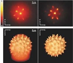
To illustrate the technique's advantages, the researchers recorded images of a spiked pollen grain 30 µm in size. With no time multiplexing, the x-y image recorded by a MMM having foci separated by 5.12 µm shows significant haze (top left); with multiplexing added, the haze disappears (top right). Reconstructed three-dimensional (3-D) images of the pollen grain further highlight the improvement in quality: the 3-D image without time multiplexing has less definition (bottom left) than the multiplexed image (bottom right). The researchers believe the interfocal distance of such a MMM can eventually be reduced to the point that lateral scanning will no longer be needed.
About the Author
John Wallace
Senior Technical Editor (1998-2022)
John Wallace was with Laser Focus World for nearly 25 years, retiring in late June 2022. He obtained a bachelor's degree in mechanical engineering and physics at Rutgers University and a master's in optical engineering at the University of Rochester. Before becoming an editor, John worked as an engineer at RCA, Exxon, Eastman Kodak, and GCA Corporation.
