Ion irradiation controls metallic clustering
French physics researchers and glassmakers have collaborated to develop a "photographic" method for growing metal nanoclusters in silicon dioxide glass, which may eventually prove useful in controlling potential optoelectronic functions such as all-optical switching and filtering.
Historically, metallic nanoclusters sized on the order of a wavelength of light have occurred naturally in glass as an outcome of the glass-making process. The Mie scattering (described by Gustav Mie in 1907) caused by those nanoclusters provides much of the mystical coloration in the stained-glass windows of medieval European cathedrals.
Currently, however, the need to study potentially important optoelectronic parameters such as large third-order susceptibility, second harmonic generation, and picosecond response time has generated a corresponding need to create metallic nanoclusters in glass in a predictable and controlled fashion.
Researchers at the Universities of Orsay and Paris, at the CNRS-St. Gobain laboratory (Aubervilliers, France) and at Corning S.A. (Avon, France) have achieved this predictability in the laboratory using a process that they have dubbed ion beam "photography." The first step in the process (analogous to a photographic exposure) involved 11.9-MeV bromine, 7-MeV silicon, or 6-MeV oxygen ion-irradiation of copper oxide containing glass samples at room temperature using the ARAMIS particle accelerator (Orsay University, France). Ion fluences during irradiation were low (less than 4 x 10¹³ ions/cm2), while the ion beam flux was kept low enough to minimize temperature increases. The exposure process effectively seeded the glass sample with an entirely predictable density of pure copper cluster nuclei that depended upon the ion mass-energy combination and ion fluence during irradiation.
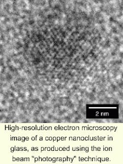
The second step in the process (analogous to the development of a photographic exposure) consisted of post-irradiation annealing of the sample at 673 K for periods of at least 600 seconds, which served to grow the nuclei into clusters of uniform size that could be observed using high-resolution electron microscopy. The researchers achieved an average cluster size ranging from 2 to 8 nm with a size distribution on the order of 2 nm full-width half-maximum. By decoupling the nucleation and growth steps (ion irradiation controlled cluster density, while thermal annealing controlled cluster-size distribution), the "photographic" process enabled the researchers to exert total control over cluster density, average size, and size distribution, which had not been achieved previously. Cluster growth during thermal annealing and its subsequent effect upon cluster size distribution corresponded to the Lifshitz-Slyozov-Wagner theory of diffusion-limited precipitate ripening (see figure).
"The analogy with a photographic process is rather striking," researchers wrote.1 "Understanding how far it may be carried requires further scrutiny of the relationship between the energy-deposition mechanism and metal precipitation." Subsequent research also has combined this process with masking techniques, with an eye toward optoelectronic and magnetic applications.
REFERENCE
- E. Valentin et al., Phys. Rev. Lett. 86, 99 (January 1, 2001).
About the Author
Hassaun A. Jones-Bey
Senior Editor and Freelance Writer
Hassaun A. Jones-Bey was a senior editor and then freelance writer for Laser Focus World.