Age-related macular degeneration (AMD) is the leading cause of blindness in the elderly in the United States. Even when not causing complete blindness, the degeneration produces a gradual reduction in visual acuity that can result in the need to use powerful magnifying glasses to recognize even a single word of text at a time. Lutein and zeaxanthin, two carotenoid antioxidant pigments that occur naturally in the retina, may help prevent AMD by scavenging free radicals produced by light, oxygen, and highly unsaturated lipids; their yellowish color may also filter out some damaging short-wavelength light. Autopsies show that carotenoid pigment levels are lower in patients with AMD.
The usual noninvasive method of measuring pigment levels is to image a flickering spot of light on the fovea and another just outside the fovea; the subject must then describe differences in colors between the two spots. Eye quality must be good and the subject cooperative for this method to work; many patients at risk for AMD may not meet either of these requirements. An objective, noninvasive method for measuring carotenoid pigment levels in the macula is needed.
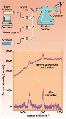
A group at the University of Utah (Salt Lake City, UT) has developed such a method, although it is still in the early stages of research. A 488-nm laser beam from an argon-ion laser is focused on the retina at an intensity level and dose falling below safety limits (see figure). The laser and optics are interfaced with a fundus camera, which permits visual identification of the macular region. The returning light is routed to a spectrograph, where its Raman spectrum is measured. A collinear beam from a helium-neon (HeNe) laser produces a red spot on the retina that serves as a locator spot for the observer viewing the subject's macula through the fundus camera.
A Raman spectrum from the macula of a human volunteer (with dilated pupils and no AMD) reveals carotenoid Raman signals atop a broadband fluorescence background. The laser dose delivered to the macula was approximately 38 mJ/cm2, a factor of 400 less than the maximum permissible safe dose. By repeating the measurement, summing the results, then subtracting out the broadband background, the signal-to-noise ratio is increased. When compared to the height of a carbon double-bond spectral peak, the height of the Raman peak shows the concentration of carotenoid in the macula. The researchers expect their instrument to be able to measure carotenoid concentrations at least a factor of 10 lower than existing techniques can measure on living human subjects. Such an ability will open up research in the prevention and treatment of AMD; indeed, clinical trials with more subjects are already under way.
About the Author
John Wallace
Senior Technical Editor (1998-2022)
John Wallace was with Laser Focus World for nearly 25 years, retiring in late June 2022. He obtained a bachelor's degree in mechanical engineering and physics at Rutgers University and a master's in optical engineering at the University of Rochester. Before becoming an editor, John worked as an engineer at RCA, Exxon, Eastman Kodak, and GCA Corporation.
