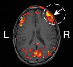
Researchers at the University of Illinois (UI; Champaign, IL) are exploring the use of frequency-domain near-infrared spectroscopy (NIRS) as a noninvasive diagnostic tool to study changes in human brain activity. In one experiment, they demonstrated a correlation between hemodynamic (blood-flow-related) signals at the surface of the brain obtained with both NIRS and functional magnetic resonance imaging (MRI)—the standard technique used in brain activation studies. Based on the results, UI physics professor Enrico Gratton believes NIRS shows promise as a way to study brain activity without performing surgery on the skull (see figure). As another benefit, the technique is both simpler to use and less expensive than functional MRI and other noninvasive methods such as positron emission tomography.
"Whenever a region of the brain is activated—directing movement in a finger, for example—that part of the brain uses more oxygen," said Gratton. "Our technique works by measuring the blood flow and oxygen consumption in the brain." The measurements are done with a two-wavelength (758 and 830 nm) frequency-domain (110-MHz modulation) oximeter from ISS Inc. (Champaign, IL). In this case, the oximeter included 16 laser diodes, eight for each wavelength, and two photomultiplier tube detectors.
Testing involved attaching an optical sensor to the left or right side of a test subject's forehead above the eyebrow and sinuses, with the detector line parallel to the eyebrow. Optical fibers carried the light from the laser diodes to the subject's forehead, where the light then penetrated the skull. The resulting scattered light was collected by optical fibers, sent to detectors, and analyzed by a computer. For the test subjects that exhibited activation signals under the nonhairy area of the forehead, measurements were repeated with the optical sensor attached to the location on the forehead where the preliminary signal indicated functional activity was occurring.
By examining how much of the light is scattered and how much is absorbed, Gratton and his colleagues in the university's Laboratory for Fluorescence Dynamics can map portions of the brain and extract information about its activity. "The degree of scattering also helps us determine where the neurons are firing," he added. "This means we can simultaneously detect both blood perfusion and neural activity."
The technique has potential uses in a variety of diagnostic, prognostic, and clinical applications. "For example, it could be used to find hematomas in children, or to study blood flow in the brain during sleep apnea," said Gratton. "It could also be used to monitor recovering stroke patients on a daily or even hourly basis—something that would be impractical to do with MRI."
To validate the technique, Gratton and Vladislav Toronov, a researcher at the university's Beckman Institute for Advanced Science and Technology, compared hemoglobin oxygen concentrations in the brain using signals obtained simultaneously by NIRS and functional MRI. "Both methods were used to generate functional maps of the brain's motor cortex during a periodic sequence of stimulation by finger motion and rest," explained Gratton. "We demonstrated spatial congruence between the hemoglobin signal and the MRI signal in the motor cortex related to finger movement."
In the research project supported by the National Institutes of Health, the researchers also demonstrated collocation between hemoglobin oxygen levels and changes in scattering due to brain activities. "By having a volunteer move different fingers, we could see an increase in perfusion in different areas of the brain," Gratton reported. "The changes in scattering associated with fast neuron signals came from exactly the same locations."
About the Author
Paula Noaker Powell
Senior Editor, Laser Focus World
Paula Noaker Powell was a senior editor for Laser Focus World.
