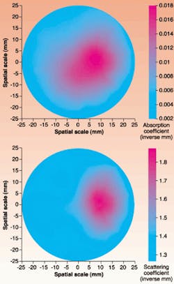
Absorption and scattering images of in vitro and in vivo bones and joints have been captured from continuous-wave tomographic measurements by researchers from Clemson University (Clemson, SC) and Medical University of South Carolina (Charleston, SC), in what is believed to be the first demonstration of diffuse optical tomography's (DOT) potential to allow early detection and monitoring of bone and joint diseases.
Normal and diseased bone/joint tissues show clear differences in absorption and scattering coefficients. The idea behind using DOT to determine bone/joint health is to extract spatial maps of intrinsic tissue absorption and scattering coefficients from measured optical data through model-based reconstruction methods, reports the research team. Bone and joint diseases affect millions of people worldwide. "Existing techniques such as x-ray or ultrasound only provide tissue structural information," explains Huabei Jiang, an assistant professor in the Physics and Astronomy department at Clemson University. "These techniques are not appropriate for detecting early changes of these diseases. Diffuse optical tomography can overcome these limitations, and is also suitable for monitoring these progressive diseases."
To test the technique, in vitro and in vivo optical data was collected for human finger bones and chicken bones embedded in a cylindrical scattering media. The bones were imaged at multiple transverse planes with a multichannel diffuse optical imager. Intensity-modulated light from a 785-nm, 50-mW diode laser was sent to bone samples in 16 3-mm fiberoptic bundles. For each source position, the diffused light was received at 16 detector positions along the surface of the sample and delivered to a photomultiplier tube (PMT). A second PMT recorded the reference signal. Direct-current data was obtained and recorded with reconstruction software to create a two-dimensional, cross-sectional image of the medium/tissue at the source-detector plane.
In the in vitro experiment, the sample consisted of two chicken bones embedded in a cylindrical solid phantom with a diameter of 5 cm. The phantom contained a scattering medium composed of a fat emulsion suspension, with India ink as an absorber and Agar powders to solidify the fat emulsion/ink suspensions. Measurements were recorded at two transverse planes, and the absorption and scattering images were recovered from the measurements with a nonlinear, finite-element-based reconstruction algorithm. These images show a marked absorption or scattering increase at the bone region, compared to the phantom background. The bone size resolved by DOT was 1.50 cm, which is comparable to the bone sample's actual size of 1.60 cm. This size was determined based on the full width at half maximum of the recovered bone optical properties. Average bone absorption and scattering coefficients were calculated to be 0.021/mm and 1.75/mm, respectively.
In the in vivo experiment, a male volunteer's index finger was used as the sample, and inserted into the same cylindrical solid phantom and scattering medium used in the in vitro experiment. Measurements were recorded at the joint and off-joint/bone planes. Both absorption and scattering images of the sample were recovered from the measurements with the reconstruction algorithm. These images show a significant absorption and scattering increase in the joint and off-joint bone regions related to the surrounding soft tissue and phantom background (see figure). The size of the bone sample imaged was 1.65 cm, which is comparable to the actual bone size of 1.70 cm. Average bone absorption and reduced scattering coefficients were calculated to be 0.023/mm and 2.14/mm, respectively. A joint containing synovial membrane and fluid showed significantly smaller absorption and scattering coefficients than the bone region, which were determined to be 0.011/mm and 1.81, respectively.
The research team believes that DOT may become a powerful tool for detecting and monitoring bone and joint diseases such as arthritis and osteoporosis, primarily because the method is capable of obtaining tissue-function information and a high image resolution not possible with conventional imaging techniques.
REFERENCE
- Y. Xu et al., Opt. Exp. 8, 447 (March 26, 2001).
About the Author
Sally Cole Johnson
Editor in Chief
Sally Cole Johnson, Laser Focus World’s editor in chief, is a science and technology journalist who specializes in physics and semiconductors.
