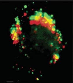
The progress of fluorescent semiconductor nanoprobes crossing a cell membrane is depicted in this image consisting of green and a red exposures taken four minutes apart (the yellow areas result from the overlap of green and red). The nanoprobes are based on cadmium selenide and cadmium sulfide quantum-dot technology developed by Paul Alivisatos and colleagues at the Lawrence Berkeley National Lab (LBNL; Berkeley, CA) Materials Science Division. The quantum dots are used as fluorescent probes; the researchers have licensed the technology to Quantum Dot (Hayward, CA) for use in biological assays.
Motivated by the possibility of designing intracellular probes with lifetimes that might exceed the lifetimes of conventional labels by two to three orders of magnitude, Fanqing Chen, in the LBNL Life Sciences Division, and Daniele Gerion, at Lawrence Livermore National Laboratory (LLNL; Livermore, CA), worked with the Alivisatos team for four years to design a stable and nontoxic probe that could be transported and subsequently operated within a living cell.1
“We took the tool Paul developed and applied it to a problem faced by biologists every day-getting inside the nucleus, a desirable target because the cell’s genetic information resides there,” Chen said.
To fabricate a probe small enough to pass through the 20-nm-wide pores in a cell membrane, the researchers designed a compact cadmium selenide/zinc sulfide quantum dot coated with silica. They also attached a protein, obtained from the SV40 virus, to the quantum dot. The protein binds to the cell’s nuclear trafficking mechanism, allowing its host to enter unnoticed. “We knew we could get quantum dots inside a cell, but getting them through the nuclear membrane is very difficult,” Chen said. “So we learned from the virus.”
To date, Chen and Gerion have introduced and retained quantum-dot nanoprobes in living cell nuclei for up to a week, and the probes fluoresce for days at a resolution high enough to detect biological events carried out by single molecules. Conventional probes, such as organic fluorescent dyes and green fluorescent proteins, fluoresce at high resolution for only a few minutes. “Our work represents the first time a biologist can image long-term phenomena within the nuclei of living cells,” Chen said.
The ability to visualize the passage of their nanoprobes across the nuclear membrane should now enable the researchers to study nuclear trafficking mechanisms in real time. Future goals include tailoring the quantum dots to track specific chemical reactions within cell nuclei. And, beyond the cell nucleus, the nanoprobes may also find use in studying organelles such as mitochondria and Golgi bodies, as well as the transfer of material between cells. “We can have two different quantum dots in two different cells, and watch as they exchange their mitochondria,” Chen said.
REFERENCE
1. F. Chen, D. Gerion, Nano Letters 4(10) 1827 (2004).
About the Author
Hassaun A. Jones-Bey
Senior Editor and Freelance Writer
Hassaun A. Jones-Bey was a senior editor and then freelance writer for Laser Focus World.
