Advanced Imaging/Fluorescence: Macro-FLIM provides molecular specificity with cellular resolution
Having overcome important limitations of fluorescence intensity imaging, fluorescence lifetime imaging (FLIM) and other time-resolved methods are increasingly recognized as beneficial for cell microscopy. As time-resolved imaging pioneer Laura Marcu discussed in a recent webcast, FLIM is currently undergoing exciting development, including progression toward clinical application.1 Now, a multidisciplinary team has developed a confocal macro-FLIM approach that not only extends the analysis area from millimeters to 4 cm2, but is the first to simultaneously provide good spatial resolution and high molecular specificity.2
The macro-FLIM approach is valuable for its ability to enable not only acquisition of structural information, but also observation of certain biochemical processes in vivo. It enables probing of biological processes, making it a tool to elucidate disease development and monitor responses to therapies. With further work, the method could even distinguish tumor boundaries during surgery with great sensitivity and precision.
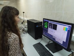
The team, including engineers and physicists from Becker & Hickl (Berlin, Germany) and biologists from Privolzhskiy Research Medical University (Nizhny Novgorod, Russia), demonstrated their macro-FLIM system by observing metabolic processes inside a whole tumor in a living mouse—an accomplishment not possible with current FLIM systems.
FLIM and macro-FLIM
FLIM works by precisely measuring the fluorescence decay of a molecule that is either intrinsically fluorescent or tagged with a probe. Because decay rate, or lifetime, is affected by such characteristics as temperature, pH, and interaction with neighboring molecules, FLIM can provide information about a tissue’s microenvironment.
Typically, laser scanning confocal microscopy is used to perform FLIM. To extend the technique to the macroscale, the researchers used picosecond lasers, high-sensitivity detectors, and time-correlated single-photon-counting (TCSPC) electronics. Placing specimens in the intermediate image plane allowed visualization of samples larger than standard FLIM systems can accommodate.
To demonstrate cellular resolution, they used the macro-FLIM system to image 14.6 μm fluorescent microbeads as well as fluorescently labeled live cultured cancer cells. And they demonstrated in vivo utility by using it to analyze an entire tumor in a living mouse: They simultaneously measured the fluorescence lifetime of both a genetically encoded red fluorescent protein identifying the tumor’s location and a low quantum yield fluorophore called nicotinamid adenine dinucleotide (NADH). The system provided sensitivity sufficient to observe intrinsic fluorescence of NADH and other tissue components without labeling. And the technique promises to enable cellular-resolution study of a tumor’s oxygen status, for instance, on a macroscale.
The work is noteworthy because most whole-body imaging approaches provide fast image acquisition, but no molecular sensitivity and poor spatial resolution. And while various efforts have been successful in addressing certain aspects of these limitations, none have overcome both.
Next steps
To develop the system for clinical applications, the researchers are working to improve its flexibility and mobility. They also want to combine the macro-FLIM system with a scanning stage that would move the sample to allow FLIM to be performed on areas as large as 10 × 10 cm.
In addition to biological and clinical samples, the macro-FLIM could be used to analyze other samples with large areas. For example, it could offer a non-destructive method for determining the media used in paintings that need restoring.
REFERENCES
1. L. Marcu, “Solving problems with time-resolved fluorescence” (2018); https://goo.gl/RAuWpD.
2. V. I. Shcheslavskiy et al., Opt. Lett., 43, 13, 3152–3155 (2018); doi:10.1364/ol.43.003152.
About the Author

Barbara Gefvert
Editor-in-Chief, BioOptics World (2008-2020)
Barbara G. Gefvert has been a science and technology editor and writer since 1987, and served as editor in chief on multiple publications, including Sensors magazine for nearly a decade.
