High-Speed Imaging: High-speed images capture processes in botanical systems
DWIGHT WHITAKER
The first example of using a series of rapidly taken images to analyze the biomechanics of a living organism goes back to the work of Eadweard Muybridge in 1878, a full decade before the first motion picture was made. In this famous experiment sponsored by Leland Stanford, Muybridge used a collection of 12 cameras triggered in rapid succession to analyze a horse’s gallop. The sequence of 12 images demonstrated clearly that indeed all four hoofs of the horse simultaneously come off the ground during each stride.
Today the technology of high-speed imaging photography has progressed to a point that digital cameras such as the Redlake (Tallahassee, FL) HG-XL can record images with resolutions exceeding 1.6 megapixel at frame rates of up to 1000 frames per second (fps) or record images with a 129 × 9 resolution at rates up to 1 million fps using the Vision Research (Wayne, NJ) Phantom V12. Such systems have enabled researchers to analyze ultrafast biological movements with unprecedented spatial and temporal resolution.
Image-quality tradeoffs
New camera systems are able to compensate for the short exposure times associated with high frame rates by using sensitive detectors. Because the maximum exposure time for each frame of a video is the inverse of the frame rate, lighting must be provided for exposure times faster than 100 μs to record images at high speeds such as 10,000 fps. To increase the sensitivity of charge-coupled-device (CCD) arrays, manufacturers have borrowed a long-standing trick of astronomers and increased pixel sizes. The tradeoff that comes from shifting to these larger pixel sizes is that the CCD array for a high-resolution camera becomes so large that standard video camera lenses are not suitable for providing distortion-free images over the whole chip. Consequently, cameras with the highest resolution sensors typically have less light sensitivity, and with nearly all systems one must carefully choose a lens suitable for the larger array sizes.
The sensitivity of a digital camera is characterized by its ISO number, which is virtually equivalent to the now obsolete ASA rating of film speed. Typical values for the ISO are between 600 and 1000 for color imaging systems and between 1500 and 3000 for monochromatic systems. These values can be compared to typical values for motion-picture film stock, which vary from 50 to 500. This added sensitivity is particularly important for biological applications as opposed to many industrial ones because simply throwing more light on your subject can cause damage to tissue.
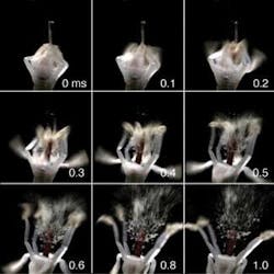
A significant challenge in obtaining good high-speed videos of biological systems is to provide sufficient lighting for the short exposure times associated with high frame rates. The high frame rates and correspondingly short exposure times, as well as the superior sensitivities of modern digital CCD arrays, have made it possible for light levels to remain low enough—even at frame rates exceeding 10,000 fps—to adequately illuminate plants without noticeable thermal damage. Our group is looking at the dynamics of the spore discharge of Sphagnum moss (Fig. 1). To get at the details of the hydrodynamics of these explosions we require images of the initial explosion at very high frame rates (10,000 to 100,000 fps) to measure acceleration of the spores and to see how the flow fields are initiated; we need images at lower frame rates (1000 fps) to understand how these flow fields govern the motion of the spores once they are released.
To achieve such rapid frame rates for sensitive biological systems, great care was taken to control the spectrum of the illuminating light source. It is crucial not to flood the system with infrared radiation that heats up the sample, but is not picked up by the camera. To record at the highest frame rates, we used fiber-coupled light sources and discharge vapor lamps as opposed to incandescent light sources. High-powered light-emitting-diode (LED) arrays have also become available with a spectrum tailored to the camera’s frequency response. These sources are also favorable because they are powered by direct current and do not produce the flicker perceivable in light sources powered by alternating current. Additionally these LEDs can be turned on and off very quickly and can even be synchronized to flash only when the camera’s electronic “shutter” is open, thus lowering unnecessary heat input when the camera is not recording data.
In addition to the limitation of extremely short exposure times at high frame rates there is also a tradeoff between the resolution of images and frame rate. Because the light level for each pixel is proportional to the number of charges accumulated within it, the overall frame rate of the camera depends on the rate at which the charges can be measured divided by the number of pixels per frame. Modern systems can read more than six billion pixels per second, limiting a 1-megapixel video to only about 6000 fps. For cameras with higher resolution the full-resolution frame rate is lower, with most systems capable of providing full resolution images at up to 1000 fps. Higher frame rates are achieved by exposing and recording only a portion of the CCD, though the resolution decreases more than proportionally with the increase in frame rate. At frame rates of 10,000 fps typical resolutions are between 0.1 and 0.5 megapixel, which is usually suitable for most applications.
Because these cameras are capable of recording data from so many pixels per second, storage and transfer of data can often be problematic. All modern cameras use onboard storage to buffer data, which can then be transferred to a computer. The availability of cheap random-access memory (RAM) has made it possible for current models to store up to 32 gigabytes of data, which amounts to about 20000 frames at 1.7-megapixel resolution. While it is a great luxury to be able to record this much data, transferring it to another computer even with Gigabit Ethernet can be tedious. Hard-drive space can also be easily eaten up with only a few videos of this size. Fortunately most systems allow the user to transfer only the frames they want to the computer, reducing transfer time and minimizing wasted hard-drive space. Many imaging systems will also encode video before transferring data to the computer, producing video files that are significantly smaller than the files for individual frames.
One of the best features of digital cameras as opposed to film—particularly for biomechanical applications—is that they allow the user to trigger the camera after an event of interest. This means that one can simply wait for something to happen while recording. Cameras can be triggered via a transistor-transistor-logic pulse from an external switch or from the computer. It is even possible to change frame rates on triggering, which can be extremely useful for recording events that happen on very different time scales, provided a suitable trigger can be used.
Capturing biomechanics
The availability of affordable, digital cameras with a continuous buffer has opened up exciting new research avenues in the area of biomechanics—the high-speed movement of biological systems such as plants, fungi, and animals. Before the advent of these cameras, capturing motion at high frame rates required elaborate triggering systems and stroboscopic lighting.
Besides Muybridge’s work there were a number of beautiful experiments performed with this old technology. In two important examples—the study of the discharge of spores from Pilobolus fungi, and the seed expulsion of Arcuethobium1, 2—the strobes were triggered mechanically and optically, respectively, from the exploding particles emanating from fixed botanical objects. While the images captured could record the speeds of discharge they were unable to capture the earliest dynamics of the explosion because the lights were always triggered after it had commenced.
The limitations of stroboscopic imaging techniques are evident in the study of the spore launch of the ballistospore.3 Video taken of this spore launch shows how water moving onto the spore causes it to be launched through the release of elastic energy liberated as the surface area of the water droplet grows as it envelops the spore. Even if one were to trigger a sequence of strobe flashes after the launch of a spore, it would be impossible to use the data from these images to show how the launch was initiated. Furthermore, stroboscopic images are most useful when a moving object is traveling in a line such that images from successive flashes do not overlap. This was not the case in our study of exploding bunchberry flowers in which our video analysis of the stamen motion revealed an optimal passive launching scheme for pollen using elastic energy stored in the filaments (see Fig. 2).4, 5 Finally, it would be virtually impossible to devise a triggering scheme to record unpredictable events (such as how a predator strikes its prey).6Available research has exploded and now covers a vast array of investigations over a number of taxonomic groups from fungi and animals to plants. Recent studies that merit mention include how birds make sounds, how bats use their wings to hover, how greyhounds run, and how froghoppers leap.7-10 Ultrafast digital imaging systems enable scientists to easily perform a number of studies on the biomechanics of rapid motion that would have been previously impossible or prohibitively difficult. The results of these analyses, coupled with field observations and morphological analysis, improve understanding of the adaptive significance of these behaviors by isolating the features required for the organism to move.
REFERENCES
1. R.M. Page, Science 146, 925 (1964).
2. T.E. Hinds, F.G. Hawksworth, and W.J. McGinnies, Science 140, 1236 (1963).
3. A. Pringle et al., Mycologia 97, 866 (2005).
4. J. Edwards et al., Nature 435, 164 (2005).
5. D.L. Whitaker, L.A. Webster, and J. Edwards, Functional Ecology 21, 219 (2007).
6. S.N. Patek, W.L. Korff, and R.L. Caldwell, Nature 428, 819 (2004).
7. K.S. Bostwick and R.O. Prum, Science 309, 736 (2005).
8. F.T. Muijres et al., Science 319, 1250 (2008).
9. J.R. Usherwood and A.M. Wilson, Nature 438, 753 (2005).
10. M. Burrows, Nature 424, 509 (2003).
DWIGHT WHITAKER is assistant professor of physics at Pomona College, 610 N. College Way, Claremont, CA 91711; e-mail: [email protected]; www.pomona.edu.

