Hyperspectral fluorescence images bacterial pigments during photosynthesis
Scientists at Arizona State Unversity (ASU; Tempe, AZ) are using hyperspectral fluorescence imaging to identify and visualize discrete pigments in live bacteria cells. This approach could help researchers fine tune the bacteria for specific purposes, according to Wim Vermaas, a professor in ASU’s School of Life Sciences.

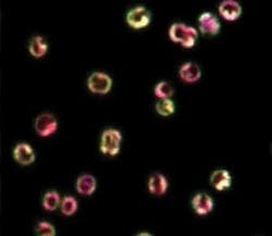
Confocal fluorescence microscopy is an excellent method for localizing pigments in cells, as long as there is little spectral overlap between different fluorescing pigments. Hyperspectral fluorescence imaging pushes the boundaries of this technique by separately localizing pigments with similar fluorescence spectra. In experiments conducted by Vermaas and colleagues from ASU’s Center for Bioenergy and Photosynthesis and Sandia National Laboratories, a 488 nm laser was used to excite phycobilins and carotenoids in cyanobacterial cells, yielding fluorescence from phycocyanin, allophycocyanin, allophycocyanin-B/terminal emitter, and chlorophyll a. Resonance-enhanced Raman signals and very weak fluorescence from carotenoids were also observed. The research team found that phycobilin emission was most intense along the periphery of the cell whereas chlorophyll fluorescence was distributed more evenly throughout the cell, suggesting that fluorescing phycobilisomes are more prevalent along the outer thylakoids. According to Vermaas, this means that even in a simple and small cyanobacterial cell there is an exquisite functional division of labor between membranes inside the cell, with different processes in photosynthesis in different areas of the membranes. Contact Wim Vermaas, [email protected].