Biophotonic imaging reveals cellular and subcellular details
The annual meeting of the Stanford University Photonics Research Center (Stanford, CA) provides an interdisciplinary academic and university showcase for leading-edge photonics research. While last year's meeting focused on recognizing the continuing importance of telecom technology within a much broader photonic landscape, the focus of this year's meeting (Sept. 15–17) was clearly on rapidly growing application areas, as well as major technological breakthroughs (in some cases as a result of telecom research efforts) in older and more traditional areas of photonic technology.
"The new areas—biophotonics and nanophotonics—are going strong, and they are offering whole new opportunities," said Robert Byer, codirector of the SPRC. "But the more classical areas of nonlinear optics are equally impressive." High points in the more classical areas included high-power fiber lasers and solid-state laser power scaling, as well as nonlinear materials and modulated-growth gallium arsenide.
Biophotonics took on a special significance at this meeting, however, as the first application-oriented technical session of the meeting and also as the subject of an industry panel session at the end of the first day. "Stanford's interdisciplinary research programs allowed us to start biophotonics a number of years ago," Byer said. "And the outcome of that has been a new facility on campus, the Clark Center for biology, medicine and engineering research in biology. It's an interdisciplinary center focused on biological systems, with the photonics application meaning the use of photonics in biological sensing."
Optical biopsy
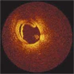
A primary focus of the biophotonics technical presentations was on nondestructive imaging and visualization methods to depict biological entities and processes at cellular and subcellular levels. James Fujimoto from the Massachusetts Institute of Technology (MIT; Cambridge, MA) described an optical-coherence-microscopy technique that obtains both high-resolution and high-depth-of-field images by combining optical-coherence tomography (OCT) with confocal microscopy. The MIT researchers are collaborating with MEMS researchers at the University of California Los Angeles and have performed cellular imaging at resolutions as fine as 1 µm. The OCT technique essentially fills the resolution niche between ultrasound and microscopy, Fujimoto said. Nondestructive "optical biopsy" capabilities have also been achieved by analyzing the composition of tissues and lesions based on spectroscopic and polarization data. The OCT technology has been commercialized through a startup, LightLab Imaging (Littleton, MA), that was purchased last year by the Goodman Co. (Nagoya, Japan), an interventional-cardiology company (see figure).
Lev Perlman from the Harvard Medical School (Cambridge, MA) described the development of an imaging method based on spectroscopy of backscattered light from cellular components, called light-scattering spectroscopy. Based on in-vitro studies, to date it is capable of imaging subcellular structures below the diffraction limit, down to about 140 nm. Georg Schuele, a graduate student in ophthalmology at Stanford, described a multi-institutional project with the Medical Laser Center (Lübeck, Germany) and Harvard University that developed two methods for noninvasively monitoring retinal temperatures during retinal laser treatments. One method involved light-scattering spectroscopy and the other involved measuring the amplitude of the optoacoustic stress waves produced by the absorption of laser pulses in tissue.
"Many of us who sat through the session hope that the opportunities lead to success, because they are health related," Byer said. The preponderance of health-related research is probably not accidental, however. In responding to a question posed to the biophotonics industry panel, Tom Baer (Arcturus; Mountain View, CA) pointed out that U.S. government funding priorities tend to make it difficult to get funding for basic biophotonic research that is not health related. The National Institutes of Health are disease focused and the National Science Foundation stays away from medicine, he said.
