LASER-TISSUE INTERACTION: Endogenous chromophores alter plasma formation during microsurgery

Laser surgery has been around for so many years it is surprising to find there are still fundamental tissue-interaction studies that have not been performed. But researchers at Vanderbilt University (Nashville, TN) recently completed a series of experiments that provide new insights into what happens when ultraviolet (UV) laser energy interacts with tissue during microsurgery, compared to the same experiments done in water.
“This is the first study that looks at the plasma dynamics of ultraviolet lasers in living tissue,” said Shane Hutson, assistant professor of physics at Vanderbilt who conducted the research with post-doctoral student Xiaoyan Ma. “The subject has been extensively studied in water and, because biological systems are overwhelmingly water by weight, you would expect it to behave in the same fashion. However, we found a surprising number of differences.”
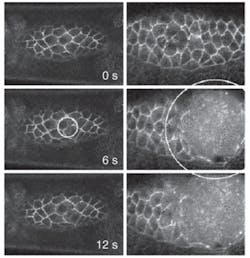
Hutson and Ma compared plasma and cavitation dynamics during laser microsurgery in water to that in living fruit fly embryos in vivo at visible and near-UV wavelengths.1 Using both the second and third harmonic (532 and 355 nm, respectively) of a Q-switched Nd:YAG laser (4 ns pulsewidth) through an inverted confocal microscope, they took fluorescent images of thick biological samples while simultaneously cutting the samples with single or multiple pulses at any user-defined location (see figure).
Hutson said his group regularly uses this system to produce UV microbeams that allow them to “tease apart” the mechanics of cell biology. By generating a near-diffraction-limited spot of UV light and adding computer-controlled beam-steering capabilities, they are able to record high-resolution, time-lapsed image sequences of the fly embryos before, during, and after UV surgery. This provides unique insight into the morphogenic process and tissue response to laser surgery, according to Hutson. For example, they have been surprised to discover that their third-harmonic results were actually quite different from those at the second harmonic, Hutson noted. In fact, they found that near-UV wavelengths work better in tissue than would be assumed based on similar experiments done in water.
“There has not been a lot of recent work for near-UV lasers and what they might be able to do in surgical applications,” Hutson said. “Far-UV lasers such as the excimer work really well in corneal tissue, but maybe in other tissues you don’t have to use the excimer, such as near-infrared solid-state lasers in the third or fourth harmonic.”
More UV surprises
Other findings also surprised them, Hutson said, such as how the elasticity of tissue changes the plasma-formation threshold. By stretching and absorbing energy, the biological matrix constrains the growth of the microexplosions. As a result, the explosions tend to be considerably smaller than they are in water. This reduces the damage that the laser beam causes while cutting flesh. This effect had been predicted, but the researchers found that it is considerably larger than expected.
Another unexpected difference involves the origination of the individual plasma bubbles. All it takes to seed such a bubble is a few free electrons. These electrons pick up energy from the laser beam and start a cascade process that produces a bubble that grows until it contains millions of quadrillions of free electrons. Subsequent collapse of this plasma bubble causes a microexplosion. In pure water, it is very difficult to get those first few electrons. Water molecules have to absorb several light photons at once before they will release any electrons.
“But in a biological system there is a ubiquitous molecule, NADH (nicotinamide adenine dinucleotide), which cells use to donate and absorb electrons, and it turns out that this molecule absorbs photons at near-UV wavelengths,” Hutson said. “So it produces seed electrons when exposed to UV laser light at very low intensities. This means that in tissue containing significant amounts of NADH, UV lasers don’t need as much power to cut effectively as people have thought.”
According to Hutson, these experiments demonstrate that the size of laser-induced cavitation bubbles is a major determinant of the region disrupted during in vivo laser microsurgery. In addition, researchers and surgeons cannot rely solely on the findings of experiments done in water to determine the optimal wavelength choice for pulsed laser microsurgery. In their next experiments, he and his group will attempt to quantify whether 405 nm is the optimum wavelength for biological research.
“It has long been generally felt in biological research community that 405 nm is the optimum wavelength for most experiments, but you can’t find anything in the literature that says this,” he said. “And people are starting to switch from the nitrogen laser to the Nd:YAG laser, so we are also going to look at the differences between the two types of systems.”
REFERENCE
1. M.S. Hutson and X. Ma, Phys. Rev. Lett. 99, 158104 (2007).
About the Author
Kathy Kincade
Contributing Editor
Kathy Kincade is the founding editor of BioOptics World and a veteran reporter on optical technologies for biomedicine. She also served as the editor-in-chief of DrBicuspid.com, a web portal for dental professionals.