Catch a butterfly in a bottle, and any chance to observe its real-life behavior is lost. The same is true for aquatic microbes, which are traditionally studied by filtering them onto a membrane that is then placed under a conventional microscope. Although a fluorescent stain makes bacteria more visible in this setup, nothing can restore their freedom
to roam and be themselves. Confocal microscopes can image three-dimensional specimens, but are too slow to keep up with moving microorganisms. Researchers at the Scripps Institution of Oceanography (La Jolla, CA) have developed a laser illuminator that, when paired with a standard microscope, lights up mobile microbes in all their glory.1
The goal was an illuminator that required no scanning, encompassed the entire focal plane of the microscope objective lens, and yet imaged only specimens that lay within the objective's focal depth. Such restrictions led the researchers to a design that projects a light sheet only microns thick through the specimen-containing solution. The 10x objective itself has a numerical aperture of 0.22, yielding a focal depth in water of 17 μm. In the illuminator, collimated light from a 488-nm-emitting argon-ion laser is expanded, sent through a 6.5-mm-diameter iris diaphragm, and focused by a cylindrical lens with a focal length of 150 mm, producing ƒ/23.08.
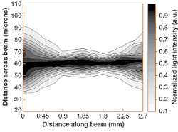
The result is a sheet of light 23 μm thick, 1 mm long, and 6.5 mm wide coinciding with the focal plane of the objective lens, and with an illumination thickness extending only very slightly beyond the objective's focal depth. The extent of the illuminated volume was measured with a fiberoptic probe mounted on translation stages; the 4-μm diameter of the fiberoptic tip was deconvolved with the experimental results to arrive at an intensity distribution (see Fig. 1).
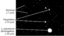
Because much of the 0.4-W laser beam goes into the light sheet, the illuminator provides strong fluorescence excitation of stained microbes. The optical power is high enough to cause local heating of the sample, so the researchers placed a shutter in the beam and synchronized it with the camera, opening the shutter for a mere 15 ms per exposure.The researchers took microphotographs of seawater in a glass cuvette, both with and without the optical system's cylindrical lens in place. When the lens is absent, the cuvette volume is flooded with light and no organisms are seen. With the lens in place, an image like a starry sky is seen, where each star is a microorganism.
In a look at unfiltered seawater, several different kinds of organisms can be seen (see Fig. 2). The sensor saturates for larger organisms, distorting their size; this effect results from the high sensitivity required to detect the fluorescence signal from smaller organisms such as bacteria.
The monochrome camera does not detect something seen by the human observer: the differentiation of natural red fluorescence of chlorophyll from artificially induced (dyed) green fluorescence of bacteria. Replacement of the camera by a color version would allow these differences to be imaged.
REFERENCE
1. Eran Fuchs et al., Optics Exp. 10(2), 145 (Jan. 28, 2002); www.opticsexpress.org.
About the Author
John Wallace
Senior Technical Editor (1998-2022)
John Wallace was with Laser Focus World for nearly 25 years, retiring in late June 2022. He obtained a bachelor's degree in mechanical engineering and physics at Rutgers University and a master's in optical engineering at the University of Rochester. Before becoming an editor, John worked as an engineer at RCA, Exxon, Eastman Kodak, and GCA Corporation.

