Double helix-PSF enables superresolution 3-D imaging
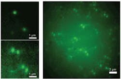
Researchers at Stanford University (Palo Alto, CA) and the University of Colorado at Boulder have demonstrated for the first time a method for 3-D optical imaging of objects smaller than 20 nm over a wide spatial range–overcoming the optical diffraction limit by an order of magnitude. Their approach uses single fluorescent molecules and a wide-field microscope. The researchers engineered the microscope’s point-spread function (PSF) to have two points of light in the image plane. Because the angle of the line between the spots changes depending on the axial position of the molecule, the PSF appears as a double-helix along the z axis of the microscope–which is why the researchers term it the double-helix PSF (DH-PSF).
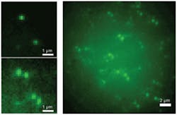
The researchers report they are able to localize single fluorescent molecules within 10 to 20 nm over a large depth of field (2 µm) in a thick polymer sample. They accomplish this by finding the orientation and center of the two DH-PSF spots using a fluorophore. Repeated 500 ms acquisition of sparse subsets enable superresolution imaging of high concentrations of single molecules in all three dimensions. The technique holds promise for improving 3-D imaging even further, the researchers say, and is applicable to materials science as well as biological and biomedical studies. Contact Rafael Piestun at [email protected].