BIOLOGICAL IMAGING: Stimulated-emission microscopy lases a trail

Biological imaging may be revolutionized with the advent of a new spectroscopic method to complement fluorescent imaging–and it relies on the fact that many nonfluorescent molecules can act as tiny independent lasers.
Fluorescence imaging has been a great boon to biologists in particular, because many of life’s molecules are naturally fluorescent, and separating their contributions spectrally in imaging applications is straightforward. Although many biochemical tricks can be used to tag fluorescent-labeling molecules to other, nonfluorescent molecules of interest, there is a tremendous amount of biology that such approaches leave in the dark.
Now, researchers from Sunney Xie’s group at Harvard University (Cambridge, MA) have tapped into molecules’ natural ability to undergo stimulated emission–in short, to lase. The approach works by using two incident lasers. Both the excitation and the “stimulating” or probe-pulse trains are made up of 200 fs pulses from a modelocked oscillator pumping two independent optical parametric oscillators.
The excitation beam, frequency-doubled in beta barium borate, was at 590 nm. The stimulating pulse train was intracavity-doubled and tunable from 550 to 700 nm, and delayed by about 300 fs relative to the excitation pulse–long enough to allow the creation, but not the decay, of excited states in the focal spot.
The detection is limited in the first instance because the excitation and probe beams cannot be focused down to molecule-size spots, so many of the incident photons pass through unabsorbed. But when excitation photons are absorbed, and probe photons drive stimulated emission, it shows up as a tiny gain in intensity in the probe beam.
“Stimulated emission is a well-known physical effect, but to realize that it can be used to detect those absorbing but not fluorescent molecules is not that straightforward, mainly because the relative signal itself is still small on its own,” says Wei Min, lead author on the research. “It needs to employ a second trick–high-frequency modulation and demodulation–to really make it useful.”
Min and his colleagues inserted an acousto-optic modulator in the stimulating beam, with a prism pair to counteract the frequency chirp the modulator introduces. The modulator was driven at 5 MHz by a square-wave-function generator, and the output of the detecting photodiode demodulated the beam with the help of a lock-in amplifier.
Quickly back to ground state
Previous techniques have tried to image nonfluorescent molecules by measuring the amount of light absorbed from single-beam measurements.1 However, that approach relies on creating excited states that have a frequently deleterious effect on their environment, because the excited molecules are significantly more reactive than in their ground state.
The new method gets around that because by design, just after excitation it drives the imaged molecules back to their ground state. With the current setup, the researchers can get a measurable signal in about 200 µs–fast enough for biological imaging if the sample is raster-scanned.2 However, using the modulation trick, the acquisition time is shot-noise limited, so a higher-repetition-rate laser could drive much higher acquisition rates.
Neither method inherently outdoes the signal-to-noise ratio of fluorescence imaging, which is easier to implement because the signal is easy to spectrally separate. But when nonfluorescing molecules are doing the dirty work, the chemistry of life becomes available for study in a way that fluorescence-based microscopies could never measure.
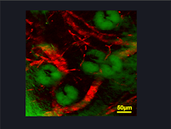
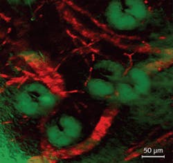
“We are working on two projects: imaging the distribution of oxygen saturation of hemoglobin across blood vessels, and imaging the oxidized or reduced states of cytochromes inside live cells,” Min says.
The method could also be used to study charge transfer in a wide variety of chemical, biological, and even physical systems. “I believe it will become a popular method for those absorbing but not fluorescent chromophores,” Min notes.
REFERENCES
- D. Fu et al., J. Biomed. Opt. 12, p. 054004 (2007)
- W. Min et al., Nature, doi: 10.1038/nature08438.
