Broadband system evaluates age and origin of bruises
A team of scientists from the Norwegian University of Science and Technology (Trondheim) has developed a simple, noninvasive optical technique that can quantitatively determine the age of a bruise. The system is expected to aid forensic scientists who previously have had to rely on more subjective methods—known as "the trained eye"— to identify how long a bruise has existed and what caused it.
"Visual inspection of the injury by a trained forensic expert is the most frequently used method," said Lise Randeberg, a graduate student at the university and lead investigator on the project. "Some experts use digital photos to aid their evaluation of the bruise. All the current methods are subjective and depend on the experience of the forensic expert. But studies show that visual evaluation of the age of bruises is very inaccurate."
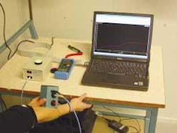
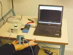
The color of a bruise depends on the depth of the hemorrhage and the chromophores present in the skin. Hemoglobin breakdown in a bruise yields several chromophores, such as biliverdin and bilirubin, which can be detected using low-cost, noninvasive optical techniques. In an effort to supplement the current visual techniques, Randeberg and her colleagues customized a commercially available fiberoptic spectrometer from Ocean Optics (Dunedin, FL) to measure diffuse reflection from the bruise and from normal skin surrounding the bruise (see Fig. 1). The spectrometer is connected to a laptop computer and uses a modified integrating sphere containing a light bulb (2800 K) to illuminate the skin with highly diffuse light and collect reflection spectra between 400 and 850 nm from the bruise and from the unbruised skin adjacent to it. The light source inside the sphere is placed behind a baffle and the light from the bulb is reflected by the walls in the sphere before it reaches the skin in front of the aperture.
"The diffuse reflectance from the skin is picked up at a small angle from the aperture to exclude specular reflection from the skin surface," Randeberg said. The signal is transmitted from the sphere to the spectrometer by an optical fiber.
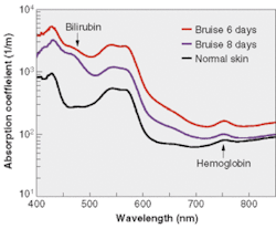
In the first pilot project, the group studied 25 bruises on 13 individuals (all sports-related injuries) and quantified the amount of bilirubin in a bruise, detected bilirubin not visible to the naked eye, and developed the parameters to be used when determining the age of a bruise (see Fig. 2).
"We are able to quantify the amount of bilirubin in a bruise and we can find bilirubin before it is visible to the naked eye," Randeberg said.
The researchers are now working to refine the algorithm for aging bruises of unknown origin and are planning a larger project that will examine bruises not just on the extremities but also in the thorax region. They are also organizing a pilot project on pigs that will involve controlled experiments with known impact and the shape of the object causing the injury, with the goal of finding a correlation between the visible injury and the cause of the injury.


