Low-resolution Raman method offers low cost and portability
Raman spectrometers can provide the detailed spectral analysis of organic systems that historically has been the hallmark of mid-infrared equipment. The main drawback is the traditional price tag for Raman equipment—even with a laser diode as the scattering source, the laser remains a major expense. This may change with recent advances in low-resolution Raman spectroscopy (LRRS). Portability of the low-resolution vibrational fingerprinting technique, coupled with low cost, may provide the impetus to take the technique out of the laboratory and into the field for real-time monitoring applications such as process quality control and environmental monitoring.
Unlike high-resolution Raman spectrometers that demand careful control of laser linewidth (adding to the cost of the excitation source), low-resolution systems relinquish resolution details in favor of emphasizing basic identifying spectral features. Like near-infrared equipment, they do not necessarily resolve all spectral features. The LRRS advantage, though, is its capability to probe organic compounds by considering the identifying vibrational bands as fundamentals instead of overtone features.
Another similarity (related to feature separation) of near-infrared and low-resolution Raman techniques is that single-wavenumber resolution is rarely required to find the fingerprint feature that will identify and quantify the sample. With the low-resolution Raman spectrometer, even if frequencies are not fully resolved, a broader-band laser source suffices.
Examining hardware
The cost advantage of LRRS thus comes from the capability to use a multimode laser diode. A LRRS prototype—built and tested at the Boston University Photonics Center (Boston, MA) under a US Army contract—includes a multimode laser diode from Power Technologies Inc. (Little Rock, AR) producing more than 300 mW-higher output than traditional single-mode Raman sources-as well as a low-resolution monochromator matched to a simple charged-couple-device (CCD) detector. Rayleigh filtering and the filtering of silica Raman signals were provided by filters inserted in the fibers near the tip of a seven-around-one bundled fiberoptic immersion probe from Visionex Inc. (Atlanta, GA).
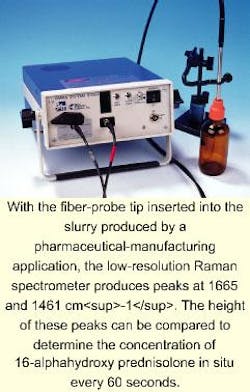
The Photonics Center also tested the resulting commercial system—the product of a joint venture between Ocean Optics Inc. (Dunedin, FL) and Boston Advanced Technologies (Boston, MA). The excitation source for the system is a 500-mW, 785-nm multimode laser diode with output delivered via a 400-µm optical fiber. The portable spectrometer measures 10 x 20 x 28 cm, weighs less than 3 kg, and, unlike traditional Raman spectrometers, costs about $10,000. For information analysis, the miniature spectrometer can interface to either a desktop or notebook PC.
The laser diode couples to a high-sensitivity spectrometer that can detect and analyze signals collected with a standard fiberoptic probe across a range of about 785-1000 nm. The detector is a linear silicon CCD array with 2048 elements—each element measures 125 x 200 µm. Well depth per element at 600 nm is 350,000 photons. Estimated sensitivity is 86 photons/count for 1-s integration. In general, integration time is 4 ms to 60 s with a 500-kHz analog-to-digital card or 20 ms to 60 s with a 100-kHz analog-to-digital card. Grating density is 1200 lines/mm, and resolution is 25 to 30 cm-1.
On the manufacturing line
A recent field test at a pharmaceutical manufacturer used the low-resolution Raman spectrometer as a real-time monitor of the manufacturing process for steroid pills. For solid oral-dosage forms and many other products in this class, it is critical to measure and control the crystalline morphology—the geometry in which the crystalline lattice is built.
Prior to development of the low-resolution Raman spectrometer, x-ray diffraction and infrared techniques were the only methods available to monitor the polymorphic state of the crystalline slurry as it is formed and grown from a solution. Both techniques require removing samples from the crystalline slurry and filtering, washing, and drying them, which can take more than an hour. During this time, there is potential to detrimentally modify the measurement of the sample`s crystal form. There also is a chance that the manufacturing process may change during this time.
To analyze the sample, scientists obtained three Raman spectra from a slurry comprised of crystalline samples of 16-alphahydroxy prednisolone hosted in the nonpolar solvent hexane. The three spectra correspond to decreasing concentrations of the 16-alphahydroxy prednisolone (200, 100, and 50 mg/ml). The ratio of the height of the alphahydroxy prednisolone peak at 1665 cm-1 to that of the aliphatic peak of the hexane host at 1461 cm-1 clearly indicates the concentration of the alphahydroxy prednisolone in the slurry. With the 2.1-mm-diameter probe tip inserted in the slurry, the low-resolution Raman spectrometer produced a fresh spectrum every 60 s.
In another application, the low-resolution Raman spectrometer was used to monitor a lamination process. During the two-part lamination of packaging films, it is critical to measure and control the blend ratio of the urethane adhesive to the curative. The ultimate performance of the lamination depends on a full and proper cure to produce full bond strength and heat-resistant properties. In many cases, pump delivery is set up at the beginning of a run or shift, but the blend ratio is not continuously monitored, so no one knows the actual blend ratio during processing. The miniature fiberoptic spectrometer obtained spectra every 60 s from a methylene bisphenyl isocyanate adhesive and a thermoplastic polymer curative mix (see figure). The peak at 1619 cm-1 indicates the concentration of the adhesive; the peak at 1731 cm-1 shows the concentration of the curative.
Environmental monitoring
Wastewater monitoring with the LRRS system was also studied with the goal of spotting cyanide contamination. In the USA alone, the gold-mining industry ingests more than 100 million pounds of cyanide annually. Other industries working with cyanide salts include electroplating, photographic development, printing, textiles, and leather manufacturing. The US Environmental Protection Agency currently requires that treated (detoxified) industrial waste water must have less than one part per million of residual cyanide before the waste can be discharged into aquatic ecosystems.
Conventional laboratory tests for cyanide detection—the Prussian-blue and the copper sulfide test—rely on detection of a color change before and after the formation of a chelating complex of cyanide. Their main problem is the poor reproducibility of results related to human error. Other options are chromatographic methods based on the same principle, but systems are relatively complex and have high installation costs. A more-recent test based on ion-selective field-effect transistors is cost-effective and fast but is vulnerable to the environment. Its requirement for an internal, stable reference electrode also impedes in situ applications.
Working with researchers at the US Army Edgewood Research, Development, and Engineering Center (Aberdeen Proving Grounds, MD), Photonics Center researchers obtained spectra from a water sample with 500 parts per billion of cyanide, first with a high-resolution Kaiser Optical Systems (Ann Arbor, MI) Holoprobe Raman spectrometer and finally with the low-resolution Raman equipment. The tests probed 1-2 ml of the water sample mixed with 2-3 ml of a commercial solution of 20-nm gold colloids and then recorded the resulting spectra. As in the other tests, measurements were taken with the tip of the immersion fiberoptic probe in the mix for 60 s. The characteristic Raman band of cyanide corresponding to the C = N stretching frequency was easy to detect with both Raman spectrometers, even though the LRRS resolution (full width at half maximum) dropped from 20 to 40 cm-1 and the signal-to-noise ratio fell from 165 to 8.
Another benefit of the LRRS approach to continuous wastewater management is the simplicity of the procedure. The entire operation, from mixing the colloid and sample to recording the spectrum, takes about 3 min.
M. EDWARD WOMBLE and RICHARD H. CLARKE are researchers at the Boston University Photonics Center, Boston, MA 02215, as well as principals of the Raman Systems Div., Boston Advanced Technologies Inc., Marlboro, MA; e-mail: [email protected]. SAMEER LONDHE, is an applications scientist at Bruker Optics, Billerica, MA. JON P. OLAFSSON is a student at Boston University.
