CONFERENCE PREVIEW: PHOTONICS WEST 2007Biomedical optics will open conference at a fast pace
HASSAUN JONES-BEY
The International Society for Optical Engineering (SPIE; Bellingham, WA) and the San Jose, CA, Housing Bureau sold out seven downtown hotels for SPIE Photonics West 2007 on the first day of registration, a record that organizers believe presages record attendance in excess of 15,000 visitors to take in 1000 exhibiting companies, 2800 technical presentations and 75 courses.
Photonics West 2007 kicks off at 8:30 a.m. Saturday morning, Jan. 20, 2007, with refresher courses and biomedical optics (BiOS) technical sessions. The two-day BiOS commercial exhibit opens at 1 p.m. and BiOS Hot Topics will be presented from 7 to 9:30 p.m. BiOS technical presentations span a gamut that includes biophotonic imaging, screening, and diagnostic technologies, as well as promising advances in biophotonic treatment methods.
In a session on multimodality imaging beginning at 1:30 p.m. on Saturday, four papers deal with optical imaging techniques applied in human studies. The novel optical techniques are used to complement established medical imaging modalities (such as x-ray and MRI) in diagnosing cancer and providing personalized approaches to therapy. They are essentially bridging the gap between established medical imaging modalities and the promise of molecular imaging.
One of these papers (6431-11), an exploratory pilot study presented by Catherine Klifa from the University of California-San Francisco, combines magnetic-resonance imaging (MRI) and diffuse optical spectroscopy (DOS) to predict breast-tissue response to radiation therapy for cancer. The researchers found that several DOS and MRI parameters appear promising for distinguishing spatially dependent and radiation-dose-dependent changes in the fibroglandular stroma of breast-cancer patients.
Another paper (6431-14) in the same session, given by Mario Khayat of ART Advanced Research Technologies (Saint-Laurent, Quebec, Canada), presents tissue-characterization results from a multi-site clinical study targeting optical tomography as an adjunct to conventional mammography. Also presented is the methodology used to translate the optical data to a more familiar x-ray paradigm.
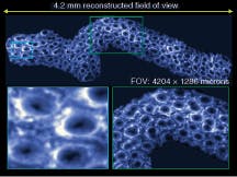
In paper 6431-13, Sacha Loiseau of Mauna Kea Technologies (Paris, France) describes how endomicroscopy has opened completely new possibilities for in situ assessment of tissue properties and pathologies. Solutions that were limited to tabletop ex vivo observations, such as multilabeling, fluorescence and autofluorescence microscopy, reflectance, and spectroscopy, may now be available as point-of-care modalities (see Fig. 1).
Gultekin Gulsen from the University of California (Irvine) describes the design of a hybrid magnetic-resonance-diffuse-optical-tomography (MR-DOT) system for dynamic imaging of cancer. The combined system acquires MR and optical data simultaneously in vivo. The group also presents preliminary results of a limited breast-cancer study undertaken using its hybrid dynamic MR-DOT imager.
Starting at 3:40 p.m. the same afternoon, a session on low-coherence light scattering, a highly promising area for medical diagnostic procedures, includes an invited talk (6446-06) by Young Kim from Northwestern University (Evanston, IL) describing a novel light-scattering modality that may change the manner in which patients are screened for colon cancer. A second invited talk (6446-07) by John Pyhtila from Duke University (Durham, NC) presents a new optical biopsy method based on light scattering that can uncover invisible precancerous lesions in the esophagus. A third talk (6446-08) describes a new method for processing optical-computed-tomography (OCT) images that may exert a significant impact on the entire field of OCT.
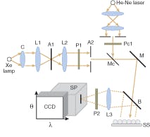
Kim’s research group at Northwestern uses a low-coherence enhanced backscattering (LEBS) to overcome problems encountered in human tissue when using enhanced backscattering techniques, which have proven useful in disciplines of physics (see Fig. 2). The group reports that, in animal studies, LEBS can detect colon carcinogenesis far earlier than is feasible by means of any conventional methods, according to Young. The researchers have also confirmed their results in an alternate animal model of colon cancer and in pilot human studies.
At 1:20 on Saturday afternoon, during a session on blood- and lymph-flow complex dynamics, Vladimir Zharov from the University of Arkansas for Medical Sciences (Little Rock, AR) will discuss in vivo laser and optical cytometry, a new approach in cytometry that allows for monitoring and quantitative estimation of parameters of blood and lymph cells moving through living organisms (6436-12).
“We believe that we have demonstrated for the first time the capability of this new technique to detect single metastatic cancer cells in vivo circulating among 107 blood cells, and to detect migrating cancer cells in lymph flow,” Zharov said. “The typical concentration of metastatic cells in blood flow is around 1:106 blood cells, and no other methods offer a potential to provide this level of sensitivity.”
On Sunday, Jan. 21, at 8:30 a. m., in a conference on microshells and nanoparticles for thermal therapy enhancement, researchers from Dartmouth-Hitchcock Medical Center (Lebanon, NH) present a paper (6440-19) on the use of iron nanoparticles as a specific cell-targeting mechanism for breast-cancer therapy. At 8:50 a.m. on the same morning in a session on clinical photodynamic therapy, Merrill Biel from the University of Minnesota (Minneapolis) presents a paper (6427-16) describing the use of photodynamic therapy for dealing with a variety of head and neck tumors that are otherwise difficult to manage.
At 2 p.m. on Sunday in a conference on endoscopic microscopy, a Stanford University (Palo Alto, CA) paper (6432-13) describes the first time that molecule-specific (peptide) probes have been administered in human subjects using a confocal microendoscope and demonstrated to bind to premalignant tissue. This result opens up a new frontier for in vivo optical imaging that targets molecules associated with disease, and may become a very powerful tool for the early detection of cancer.
Micro and nano
The BiOS exhibit ends on Sunday at 4 p. m., and the three-day Photonics West commercial exhibit begins on Tuesday at 10 a.m. The MOEMS-MEMS plenary session on Monday at 9 a.m. will include a discussion lead by Henry Smith from the Massachusetts Institute of Technology (Cambridge, MA) on the unique challenges in fabricating micro- and nanophotonic structures, a talk entitled “MEMS & Optics: A Happy Marriage” by Richard Payne from Pixtronix (Andover, MA), and a discussion of MEMS for medical technology applications with Göran Stemme, from the Royal Institute of Technology (Stockholm, Sweden).
On Wednesday in a session on display applications that begins at 11 a.m., a paper (6466-9) by Michael Scholles from the Fraunhofer Institute for Photonic Microsystems (Dresden, Germany) describes a breakthrough in the miniaturization of laser displays. The researchers used a micro scanning mirror to develop a projector the size of a sugar cube. With potential applications in the fields of automotive head-up displays, wearable displays and even projection displays for mobile phones, the technology is of extremely high interest for both public and industry. Both monochrome and full-color systems are available. All systems have VGA (640 × 480 pixels) resolution and operate with 8-bit color depth per pixel and 50 frames per second.
On Tuesday at 3:45 p.m., a session on MEMS reliability within Conference 6463, “Reliability, Packaging, Testing and Characterization of MEMS/MOEMS,” concludes with a 4:25 p.m. panel discussion on MEMS reliability. The panel is moderated by Jason Clevenger (Exponent; Menlo Park, CA) and panelists includeMEMS-reliability professionals from the Sandia National Labs (Albuquerque, NM), Analog Devices (Norwood, MA), Colibrys (Neuchâtel, Switzerland), Akustica (Pittsburgh, PA), and Qualcomm (San Diego, CA). Reliability is critical to success of MEMS products, because time to market and profitability are faster when MEMS reliability is understood and addressed, starting from the design phase and continuing through product introduction
This year’s Integrated Optoelectronic Devices (OPTO) plenary session begins at 8:30 a.m. on Tuesday and will include a discussion of transformative advances in electro-optic and all-optical materials and devices by Larry Dalton from the University of Washington (Seattle) as well as a talk on optofluidics by Demetri Psaltis from the California Institute of Technology in Pasadena, CA (see “Optofluidics reinvents the microscope,” p. 83).
Technical papers of note in the OPTO conference include a National Security Agency (Fort Meade, MD)-sponsored optical-interconnect project that has led to demonstration, with an aggregated bandwidth of 2.4 Gbit/s, of the first 16-channel optically interconnected computing system based on photopolymer-based waveguide holograms (see Fig. 3). The ultimate goal is to reach 1 terabit/s. The unit has been delivered to the National Security Agency for further evaluation. This paper (6478-02) is presented by researchers from the University of Texas at Austin.
In tune with the excitement over the development of silicon lasers, back-to-back sessions on silicon optoelectronics will be held Wednesday (see “Two groups create electrically injected hybrid lasers on silicon,” www.laserfocusworld.com/articles/274700). The first, at 1:30 p.m., is chaired by Mario Paniccia of Intel (Santa Clara, CA; see photo, p. 63); the second, at 4:00 p.m., by Bahram Jalali from the University of California Los Angeles. The joint sessions are part of conference 6477 on silicon photonics and of 6485 on novel in-plane semiconductor lasers.
Also on Wednesday, beginning at 10:30 a.m., speakers at the Lasers and Applications in Science and Engineering (LASE) plenary include Charles Townes from the University of California, Berkeley, speaking on “The Laser-Its Origin, Development and Possible Future;” Robert Byer from Stanford University on “Lasers: Astrophysics to Particle Physics;” and Hans-Juergen Kahlert of Jenoptik Laser, Optik, Systeme (Jena, Germany) on “Optical Technologies: Engine for Innovations in Industrial Applications of Lasers.”


