Enhanced imaging, detecting possible with spectroscopy
Spectroscopy runs the gamut of biological applications—detecting molecular and/or structural composition of a sample, characterizing proteins, and even improving effectiveness of medications.
Today, spectroscopy is becoming an even stronger option in biomedical research, from COVID-19 testing to brain imaging.
COVID-19 testing
Roughly two years into the COVID-19 pandemic, social distancing, face masks, and hand sanitizer at every corner are the norm. In most places, these and other preventative-measure items are no longer in short supply. But more efficient testing is.
Laboratory testing—the polymerase chain reaction (PCR) and reverse transcription PCR (RT-PCR)—is highly accurate, according to a study by a team at the Federal University of Paraná (Brazil). In their work, published in the American Journal of Infection Control, researchers found the RT-PCR test in particular, which involves a nasopharyngeal (the upper part of the pharynx, behind the nose) swab or bronchial aspirate, correctly diagnosed COVID-19 in 97.2% of cases.
However, researchers from Nanyang Technological University (Singapore) say the RT-PCR test has “a substantial false-negative rate” based on the timing of the testing in relation to when a person was exposed to COVID-19. Such tests are also time-consuming, often taking days for results. This lacks efficiency, according to the team, noting in their study, published in ACS Nano, “population-wide surveillance of COVID-19 requires tests to be quick and accurate to minimize community transmissions.”
The potential to mitigate these challenges exists, though, thanks to a spectroscopy technique harnessed by the Nanyang Tech team.
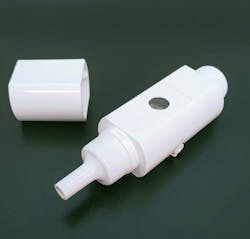
The researchers have developed a handheld breathalyzer comprised of a chip with three surface-enhanced Raman scattering (SERS) spectroscopy-based sensors attached to silver nanocubes, for screening of large groups of people. Also a benchmark for detecting lung cancer and other diseases, breath testing relies on differences in concentrations of breath volatile organic compounds exhaled by someone who has COVID-19. With the new breathalyzer, those compounds chemically interact with the SERS-based sensors.
A portable Raman spectrometer is then used to characterize the bound compounds based on key variations in vibrational fingerprints in the sensors that occur from interactions between breath metabolites and multiple molecular receptors. The researchers say “spectral regions influencing classification show strong corroboration with reported potential COVID-19 breath biomarkers” through actual experiments as well as simulations.
The smaller, easier-to-use device provides results in under 5 minutes, making it a potential alternative to even at-home rapid tests (see Fig. 1). In a study of 501 people in hospitals and airports in Singapore, the device achieved higher accuracy, with just a 3.8% rate of false-negative results and 0.1% rate of false-positives.
“This is a proof-of-concept work that demonstrates it is possible to use breath for COVID-19 detection. And this is just the beginning,” says lead researcher Xing Yi Ling, a professor and principal investigator at Nanyang Tech. She notes that more validation steps and testing will be necessary moving forward, before the device is ready for real-world application. “Our study … signifies a decisive step forward in the practical application of SERS for next-generation, point-of-care diagnostic toolkits of other respiratory and non-respiratory diseases, not limited to COVID-19.”
FAST CARS
An international team led by researchers at Texas A&M University (College Station, TX) has discovered that a blend of spectroscopy techniques can “map a single virus particle with nanometer resolution and chemical specificity.”
The combination includes spatial resolution of atomic force microscopy and the spectral resolution of coherent Raman spectroscopy to create femtosecond adaptive spectroscopic techniques for coherent anti-Stokes Raman scattering (FAST CARS) with enhanced resolution (FASTER CARS). The goal of the work, which is published in the Proceedings of the National Academy of Sciences, is to characterize a virus based on single viral particles.
The goal (in addition to virus diagnostics) is to identify structural changes of the virus surface at an early stage, using tip-enhanced Raman scattering (TERS) together with CARS.
Researchers expect the combined technique to enhance testing sensitivity and considerably speed up acquisition times. Specifically, it could allow early detection of changes “that alter the antigenic properties of viruses and thus the effectiveness of vaccines” with very small sample quantities. This makes possible the identification of a virus without specific antibodies.
Currently, PCR testing is most common in such research, as it “has simplified and accelerated the detection of pathogens over culturing techniques.” However, similar to its use in COVID-19 testing, its application in microbiological detection and diagnostics is susceptible to contamination that could prompt false-positive results. False-negative test results are also possible.
Brain imaging
Functional near-IR spectroscopy (fNIRS), which provides accurate, high-resolution observation, is already widely used for noninvasive imaging of the brain and its functions. These types of systems typically employ continuous-wave fNIRS, “where the tissue is irradiated by a constant stream of photons.” However, according to researchers at Kernel (Culver City, CA; a neurotechnology company that develops systems for studying brain function), these systems cannot differentiate between scattered and absorbed photons.
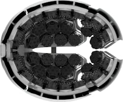
To jump this hurdle, the company is harnessing a time-domain functional near-IR spectroscopy (TD-fNIRS) technique, “which uses picosecond pulses of light and fast detectors to estimate photon scattering and absorption in tissues,” to develop a wearable whole-head headset: the Kernel Flow (see Fig. 2).TD-fNIRS systems—which emit picosecond pulses of light into tissue, enabling the measurement of single photons’ arrival times at nearby detectors—are considered the gold standard for optical brain imaging systems, the researchers say, as they produce more information than continuous-wave systems can.
The new headset system has two laser sources generating pulses under 150 ps wide. It allows coupling of the source laser light from the lasers to the scalp, the Journal of Biomedical Optics study notes, as well as the capture of return light from the scalp. It conforms to head curvature while “mitigating interference from hair, isolating detected signal between detectors, and maintaining a stable intensity at the detector.” The detector in this system, which can handle high photon counts, records the photon arrival times into histograms. The researchers collected histograms from over 2000 channels across a human brain during “a standard neuroscience validation task”—in this case, the act of finger tapping.
The system shows promise for deeper, more efficient noninvasive brain imaging, but more research is necessary before real-world use. One aspect to consider, according to Kernel CTO Ryan Field, an author of the study, will be the headset’s performance with various hair and skin types, as near-IR light is absorbed differently on each.
About the Author
Justine Murphy
Multimedia Director, Digital Infrastructure
Justine Murphy is the multimedia director for Endeavor Business Media's Digital Infrastructure Group. She is a multiple award-winning writer and editor with more 20 years of experience in newspaper publishing as well as public relations, marketing, and communications. For nearly 10 years, she has covered all facets of the optics and photonics industry as an editor, writer, web news anchor, and podcast host for an internationally reaching magazine publishing company. Her work has earned accolades from the New England Press Association as well as the SIIA/Jesse H. Neal Awards. She received a B.A. from the Massachusetts College of Liberal Arts.


