Optical design fuels innovations in intravascular imaging
Intravascular imaging gives a doctor a view inside a patient’s vascular system to help diagnose disease or other issues—and it saves lives. Cutting-edge optical technologies are helping to push innovation in this space forward with increasingly miniaturized cameras, all-optical ultrasound, and optical coherence tomography.
Tiny cameras
Miniature cameras allow doctors to see formerly inaccessible parts of the human body. An example of this is Leap from Cambridge Consultants, which features a “chip-on-tip” sensor located at the end of an endoscope, and produces a 400 x 400 pixel image of the vascular system. This improves upon traditional angioscopes greatly. Similar products are in development by other companies; Framos reports to be working on a disposable sensor that would deliver high-resolution imaging but eliminate cross-contamination or transmission of infection.
All-optical ultrasound
All-optical ultrasound is a new imaging technique where fiber-optic sensors pick up the ultrasound waves instead of today’s more common electric transducers. It enables doctors to see what they’re doing at higher resolution in real time, which has incredible implications for minimally-invasive heart or other vital surgeries.
Optical coherence tomography (OCT)
Optical coherence tomography (OCT) is a tomographic imaging system which can produce cross-sectional or three-dimensional images based on light reflected or scattered from the image. According to the American College of Cardiology, intravascular OCT is uniquely suited to detect acute coronary syndromes. “Intravascular OCT has 100 percent sensitivity (vs. 33 percent sensitivity of intravascular ultrasound) in detecting intraluminal thrombus when compared with coronary angioscopy.”
Optical design software fueling advancements
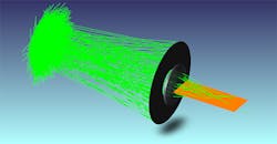
Optical design software helps optical engineers create smaller and more sensitive optical products to drive forward innovations in intravascular and many other types of medical imaging. For example, in OpticStudio from Zemax, users can model double-clad fibers, frequently used for optical coherence tomography and all-optical ultrasound systems, using non-sequential mode. This allows for definition and modeling of independent light sources of any special and angular distributions. They can also model a complete optical coherence tomography system.
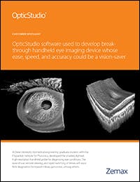
Customer story: OpticStudio used to develop break-through handheld eye imaging device
A Duke University biomedical engineering graduate student used OpticStudio to develop the smallest, lightest, high-resolution handheld probe for diagnosing eye conditions.


