SLM and software automatically focus multiple cell nuclei
Viewing a cell under a microscope can be made much easier if the cell is held in place by an optical trap (“optical tweezers”). In fact, many separate cells can be observed simultaneously, if they are all held in separate optical traps, all coplanar, created by a spatial light modulator (SLM) in the laser beam’s optical path. But even so, the cells’ nuclei will usually take up non-coplanar positions, moving many of them out of focus—a problem if the nuclei are the objects of interest.
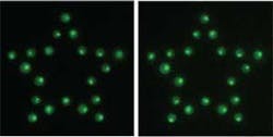
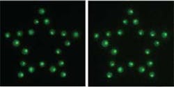
Researchers at the University of Gothenburg (Gothenburg, Sweden) and the Georgia Institute of Technology (Atlanta, GA) have used LabView software (LabView; Austin, TX) to arrive at a solution. Images of the trapped array of cells are taken through focus, and the images are filtered in software by an 8-pixel-diameter filter (about the diameter of a nucleus), finding the best focus for each cell. The SLM is then adjusted; axially shifting the cells by varying amounts to bring all the nuclei into the plane of best focus. Less-symmetric trapping patterns (such as a star rather than a grid) tend to give better performance; shown here are nuclei before (left) and after (right) adjustment. Although the researchers simply used reflected light for the optimization, they say a fluorescent stain could be used too. Contact Mattias Goksör at [email protected].

