How to select components
How to select components
for image processing
Designing a useful PC-based imaging system means integrating the
right cameras,
optics and lighting, frame grabbers,
and software to
maximize results.
Robert Gregory
Advances in bus architecture and processor speeds have made it possible to use personal-computer (PC) based imaging systems for applications unheard of just a few years ago. Although technology is quickly changing, setting up such a system is easier if some basic guidelines are considered.
Because the applications of image processing are so diverse, components can vary from system to system. However, components usually fall into several categories.
Input source and optics. The input source and optics can be a camera, microscope, scanning electron microscope, or charge-coupled-device (CCD) array. These devices are used to take a picture, or image, of the object being inspected. Depending on the application, cameras can be standard monochrome (also known as RS-170), composite color (or Y/C), red-green-blue color, nonstandard monochrome (variable scan), linescan, or custom CCD arrays (used for x-ray). Cameras can be built into a microscope or mounted separately.
Lighting. Lighting is needed to illuminate the object or specimen so that the best possible image can be acquired. Fixtures and bulbs are available in a wide variety of shapes, sizes, and intensities, but common bulb types are fluorescent, light-emitting diodes, and high-intensity lights.
X-Y table. The x-y table automates the process of acquiring images of multiple samples. The table moves a predetermined distance after each image is acquired to properly position the next object or specimen in relation to the camera.
Frame grabber. The frame grabber is a circuit board that takes image data from the camera and converts them to digital information for use by the PC. Frame grabbers are installed inside the PC chassis and come in various configurations to support different camera types (for example, monochrome, color, digital, or linescan) and computer bus platforms (for example, PCI, ISA, and VME). For PC-based image processing, the PCI bus is the most common.
PC platform. The computer is the heart of an imaging system. It is usually equipped with a 133-MHz (or faster) Pentium or Pentium II processor and Windows 95 or NT. The PC should have a minimum of 32 Mbytes of RAM, especially if processing highly detailed images.
To determine the memory needs for an application, it may help to decide how much memory each image will require. For an 8-bit monochrome image, simply multiply width by height to compute the memory needed. For example, if the image size is 1K ¥ 1K, then 1 Mbyte of memory is required for each image. If the resolution is more than 8 bits, then the formula is width by height times two.
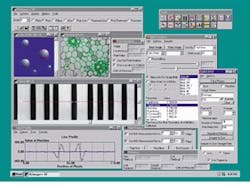
Imaging software. Software allows the image to be processed, analyzed, and stored. Different types of software packages are available, ranging from easy-to-use packages with predefined tools to software-development kits that allow programmers to build custom imaging applications (see Fig. 1).
Network connection. Once the system has acquired an image, that image and the resulting data may need to be accessed by other users. This sharing is accomplished with a network connection that links computers together, usually within an organization.
Careful planning will ensure that a PC-based imaging system meets a user`s needs. Chief among the issues to be considered in planning a system is identifying what the system is intended to accomplish. Imaging operations fall into several categories including measurement, counting, particle classification, and blob analysis. It also is important to consider how many samples will be processed immediately and in the future.
Select the right components
A PC-based imaging system is only as strong as its weakest link. Any shortcuts made in component selection, especially in the optics and imaging path, can greatly reduce its effectiveness. The basics to remember in choosing the components of the image path include the following:
Cameras. Typically, camera selection is directly tied to the application requirements. For example, a monochrome camera is usually used for general applications, while a color camera is needed for color applications. In addition, the resolution should be high enough to capture needed information. A digital camera may be the best option for the highest accuracy.
Optics and lighting. Optics and lighting are crucial considerations that are often overlooked. When poor optics or lighting are used, even the best system will not perform as well as a less capable system with good optics and adequate lighting. The right optics will produce the best and largest usable image, and correct lighting illuminates the key features being examined.
In choosing lighting for your system, first consider the other lighting in the room. Certain types of room lighting combined with the capture rate of the camera can result in artifacts on the image (often referred to as a herringbone pattern). Blocking out any outside lighting and using high-frequency fluorescent lighting will avert this problem.
Choosing lighting for microscopy applications poses a specific set of challenges. Particular attention should be paid to the illumination underneath the microscope. If the lighting is not built in, select lighting that is completely even, with a color spectrum that best illuminates the objects being examined. For applications involving live samples, avoid fixtures that generate damaging heat.
Frame grabber. Although the frame grabber may be one of the smallest parts of an inspection system, it is one of the most important because it is in the image path. Ideally, the frame grabber should convert the analog image data from the camera into digital data for use within the PC with as little alteration to the image data as possible. Choosing the wrong frame grabber can result in errors being introduced into the image data.
Software. The right software will help reap the benefits of careful hardware choices. Many manufacturers offer demonstration versions of software products that should be tried before purchase, with specific applications kept in mind.
Minimize possible problems
The human eye and brain are elaborate and versatile systems, capable of identifying objects in a wide variety of conditions, for example, identifying familiar people even when they are wearing different clothes and recognizing familiar landmarks when driving on a foggy day. A PC-based imaging system is not as versatile; it can only perform what it has been programmed to perform.
Knowing what the system can and cannot recall are important points to keep in mind to obtain desired results and reduce errors and incorrect measurements. Common variables include changes in the object`s color, changes in surrounding lighting, changes in camera focus or position, improperly mounted camera, and environmental vibration. A vibration-free environment with all extraneous light removed will eliminate many common problems.
Store and access the data
For many systems, obtaining an accurate image is only the first step. The capability to store the data and easily access it later by others is just as important. Careful planning will be needed at this stage to produce a smooth integration of the imaging system into a computer network. Common concerns that must be addressed are the types of signals required, the kind of network currently used or required, and the file formats that will be transferred. Another consideration is whether the software selected will support the networking function.
A PC-based imaging system is only as good as its weakest link and only as accurate as the information it receives. Spending some time and effort in performing the proper setup and paying attention to the details will result in an accurate and resilient system. o

FIGURE 1. Scientific imaging software for a PC provides a flexible imaging application with predefined image-processing tools.
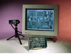
FIGURE 2. For accurate, error-free data from an image-processing system, carefully select the components in the image path. For the highest accuracy, choose a digital camera and an appropriate frame grabber.
Speeding images for cardiac patients
Patients at risk for a heart attack are usually rushed into the cardiac-catheterization laboratory. Physicians inject dye into the heart, wait for the dye to make its way through the arteries, and use results from an x-ray to diagnose blockages and other problems. Amid the labyrinth of state-of-the-art medical equipment for such procedures, the device used to record the x-ray images seems like an anomaly: a standard 35-mm motion-picture camera.
The 35-mm film has several disadvantages. First, the test cannot be viewed while it is underway, and, in fact, there is a lengthy delay while the film is developed and loaded onto a projector. Also, the procedure is expensive, averaging $100 for film alone. In addition, the film degrades over time and is bulky to store.
Fortunately, new products are providing catheterization-test results to physicians--immediately and at a significant cost savings--by replacing the film with a PC-based imaging system. For example, the Write Star system (Comview Corp.; Livermore, CA) incorporates a DT3152 variable-scan monochrome frame grabber (Data Translation; Marlboro, MA) to capture the x-ray signal during the procedure and convert it to a digital signal for processing by a computer (see figure). The PC is equipped with custom software that enables technicians and physicians to view the images from the x-ray on a monitor during the procedure, and/or archive them on a standard DICOM-format compact disk. The system also can be networked for easy access by other physicians and comparisons with previous archived tests.
R. G.
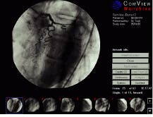
The WriteStar PC-based imaging system allows users to view cardiac-catheterization tests in real time or from an archived CD. The system uses a frame grabber to acquire x-ray images at u¥to 30 frames/s.