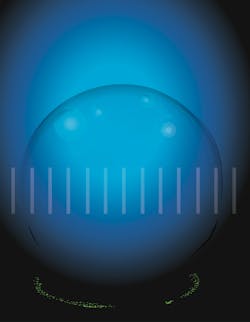
Optical microscopes capture in-flight microdroplets
National Institute of Standards and Technology (NIST; Gaithersburg, MD) researchers are improving the accuracy of optical microscopes to image in-flight microdroplets via new standards and calibrations.
It’s not easy to measure the volume, motion, and contents of microscopic droplets, which is important for studying how airborne viruses such as COVID-19 spread, the way clouds reflect sunlight to cool the Earth, or how plastic bottles fragment into nanoscale plastic particles that pollute the oceans.
Optical microscopes can directly image the positions and dimensions of small objects, so their measurements can be used to determine the volume—proportional to the diameter cubed—of spherical microdroplets. But the researchers point out that the accuracy of optical microscopy is limited by many factors, such as how well the image analysis can locate the boundary between the edge of the droplet and the surrounding space.
Beyond new standards and calibrations, the researchers also created a system to simultaneously measure the volume of in-flight microdroplets using microscopy and gravimetry. Gravimetry measures volume by weighing the total mass of microdroplets collected within a container.
If the number of droplets is controlled and the density—mass per unit volume—is measured, according to the scientists, “then the total mass registered on a scale can vary in size, imaging single droplets by optical microscopy enables a more direct and complete measurement.” They relied on gravimetry to check the reliability of microscopy to determine droplet dimensions.
The team wanted to improve the accuracy of locating the microdroplet edges, so they tested two standard objects to mimic a microdroplet and calibrate the image boundaries. For each standard object, a precisely and accurately measured distance between its edges enabled calibration of the corresponding image boundaries.
In one of these tests, the standard object was plastic spheres with calibrated diameters that produce images in the microscope very similar to those of microdroplets. And when they used the plastic spheres to calibrate their measurements of image boundaries, they found the microdroplet volume derived from microscopy precisely matched that from gravimetry.
The researchers then calibrated several aspects of the optical microscope, including focus and distortion, while maintaining the links to the International Standard of Units (SI) throughout.
After these improvements, optical microscopy resolved the volume of microdroplets to one trillionth of a liter. The researchers note that the less advanced the microscope optics are, the more a microscopy measurement can benefit from standards and calibrations to improve the accuracy of image analysis.
For their main experiment, the team used a printer to shoot a jet of microdroplets of a viscous alcohol that evaporates slowly. As the microdroplets flew into a container located a few centimeters away, they were backlit and imaged with the optical microscope. Then, the team weighed the container of microdroplets and confirmed the optical microscope was calibrated (by comparing it to the gravimetry method).
The researchers ran another experiment with water microdroplets containing nanoparticles of polystyrene, which are commonly used for nanoplastic analysis (see figure). This time, the printer deposited rows of individual water microdroplets onto a surface one at a time.
After landing on the surface, the water evaporated, leaving behind nanoparticles labeled with fluorescent dye. And the team recorded the number of particles suspended within the volume of each microdroplet, which provides a measure of concentration. This measurement, they say, is both a way to sample the bulk liquid and study properties of microdroplets containing small numbers of nanoparticles.
“Optical microscopy beyond the resolution limit presents many opportunities, with diverse applications involving localization analysis,” says NIST Researcher Samuel Stavis. “However, accurate localization is challenging to achieve across an imaging field of appreciable extent. Traceable standards for microscope calibrations are important to obtain not only precise but also accurate position data. In this way, our localization measurements of microdroplet diameters and spherical volumes reach the edge of accuracy.”
About the Author
Sally Cole Johnson
Editor in Chief
Sally Cole Johnson, Laser Focus World’s editor in chief, is a science and technology journalist who specializes in physics and semiconductors.

